

TOPIC 2: MOVEMENT AND LOCOMOTION - BIOLOGY NOTES FORM 3
MOVEMENT
Movement is the act of changing positions or postures by the part or the whole of an organism.
Movement occurs both in plants and animals
Movement in plants mainly involves growth towards or away from certain environmental factors.
LEVELS AT WHICH MOVEMENT OCCURS
Movement can occur at various levels, namely:
i. Cellular level
ii. Organ level
iii. Organism level
I. CELLULAR LEVEL
At this level, movement is by cytoplasmic streaming e.g. In amoeba and swimming of male gametes such as sperms.
II. ORGAN LEVEL
In animal, movement of organs is brought about contraction and relaxation of muscles. For example contraction of biceps muscles and relaxation of triceps muscles in human arm cause arm to be raised.
III. ORGANISM LEVEL
At this level the whole organism moves from one place to another
LOCOMOTION
Is the change in position of the whole organism from one place to another.
Locomotion occurs in animals and in some protoctists.
DIFFERENCES BETWEEN MOVEMENT AND LOCOMOTION
| MOVEMENT | LOCOMOTION | |
| 1 | It involves only part of an organism | It involves the whole organism |
| 2 | Occurs in both plants and animals | Occurs in animals and some protoctists |
IMPORTANCE OF MOVEMENT AND LOCOMOTION TO ANIMALS AND PLANTS
i. Enables organisms to escape from danger.
ii. Enables organisms to locate food and water.
iii. To move to better climatic conditions. For example birds migrate during extreme cold weather or drought.
iv. Brings together organisms or reproductive cells for reproduction.
v. Enables organisms to find good habitats
vi. To aid in insect pollination.
TYPES OF MOVEMENT
There four main types of movements, namely:-
i. Amoeboid movement
ii. Ciliary movement
iii. Flagella movement
iv. Muscular movement
I. AMOEBOID MOVEMENT
Is the movement shown by amoeba and white blood cells by using locomotory structures called pseudopodia.
II. CILIARY MOVEMENT
Is the movement shown by paramecium and larvae of some aquatic animals by locomotory structures called cilia.
III. FLAGELLA MOVEMENT
Is the movement shown by euglena, trypanosoma, some bacteria and sperm by using locomotory structures called flagella
Flagella are tail- like projection on a cell surface.
IV. MUSCULAR MOVEMENT
Is movement shown by vertebrate animals such as mammals, birds, fish and insects move by using muscles aided by skeleton.
FORMS OF LOCOMOTION
The following are various forms of locomotion exhibited by animals:-
i. Walking
ii. Running
iii. Leaping
iv. Hopping
v. Crawling
vi. Swimming
vii. Flying
I. WALKING
Is a form of locomotion shown by human beings and some animals by using two or four legs.
II. BIPEDAL
Are organisms that walk on two legs.
Example of bipedal includes;
— human being
— kangaroo
— chimpanzees
— birds
III. QUADRUPEDS
Are organisms that walk on four limbs. Example of quadrupeds includes:
— dogs
— cows — Goats
— Elephants
— zebras
IV. SWIMMING
Is a form of locomotion exhibited by aquatic animals such as fish, whales and seals by using fins and fat tissues.
V. FLYING
Is a form of locomotion shown by birds, bats and winged insects moving through air by using wings.
VI. LEAPING
Is moving by jumping from one place and landing onto another place.
à Example of leaping animals:
— Frogs
— Toads
VII. HOPPING
Is a form of locomotion shown by insects such as grasshopper moving by making quick short jumps.
VIII. CRAWLING
Is a form of locomotion shown by Earthworms, snails and millipedes moving with the body resting on the ground.
MOVEMENT OF THE HUMAN BODY
I. SKELETON
Is a rigid framework of cartilage and bones to which soft tissues, organs and muscles are attached.
OR is a framework of tissues supporting a human or an animal body.
TYPES OF SKELETON
There are three types of skeletons animals.
i. Hydrostatic skeleton
ii. Exoskeleton
iii. Endoskeleton
II. THE HYDROSTATIC SKELETON
Is a skeleton found in animals with soft bodies like earthworms.
Hydrostatic skeleton is made up of a fluid which acts as a skeleton
Hydrostatic skeleton is found in the following organisms
a. Earthworms
b. Jelly fish
c. Leech
Role of hydrostatic skeleton
Helps animals such as the earthworms to move and burrow in the soil
EXOSKELETON
Is the hard outer skeleton that covers bodies of insects and arthropods.
It is called exoskeleton because it is found outside the body of an organism.
Exoskeleton is made up by a mixture of protein and chitin.
Exoskeleton is covered with cuticle that is slippery and water proof therefore preventing loss of water from insect’s body.
Exoskeleton is made of plates called sclerites which are hard enabling insects to move.
Exoskeleton is found in the following organisms
Insects such as grasshopper, houseflies and butterflies
Arthropods such as crabs, prawns, centipedes and millipedes.
THE ENDOSKELETON
Is a skeleton which found inside the body of an organism.
Endoskeleton is made up of bone and cartilage
Endoskeleton is found in the vertebrates such:-
— Fish
— Cow
— Dog
— Human being
— Birds
— Reptiles
DIFFERENCES BETWEEN ENDOSKELETON AND EXOSKELETON
| ENDOSKELETON | EXOSKELETON |
| (i) It is found inside the body of an organism | It is found outside the body of an organism |
| (ii) It is made of bones and cartilage | It is made up of chitin |
| (iii)It is found in vertebrate animals like birds, fish and human beings. | It is found in insects like grasshoppers and other arthropods like crabs |
| (iv) it grow with the rest of the body | It does not grow because it is dead material |
| (v) it is living | It is non living |
FUNCTIONS OF SKELETON
(i) Provides the site for muscles and body organs attachment.
(ii) It protects delicate organs such as brain, heart, lungs and kidney,
(iii)It supports and gives the body its shape.
(iv) Enables the organism to move from one place to another
(v) It store minerals such as calcium and phosphorus.
(vi) Provides a rigid framework which supports softer parts of the body
(vii) Helps in the formation of blood cells such as RBCs and WBCs. E.g. endoskeleton
THE HUMAN SKELETON
The human skeleton consists of bones and joints
(I) BONE
Is a hard and tough connective tissue composed of minerals such as calcium and phosphate.
A human being has a total of 206 bones.
FUNCTIONS OF BONES
i. Provides a rigid framework which supports to the softer parts of the body.
ii. Protects the delicate organs of the body.
iii. Helps in blood cells formation in the body.
iv. Stores mineral salts in the body such as calcium.
LIGAMENT, TENDON AND CARTILAGE
LIGAMENT
Is a fibrous tissue which joins bone to bone.
Function of ligament
(i) It is elastic to allow movement at joint
(ii) It makes joints more stable.
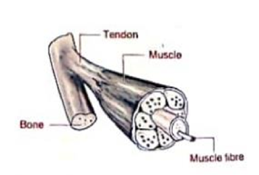
TENDON
Is a tough connective tissue that joins muscle to the bone.
FUNCTION OF TENDON
(i) It is inelastic to join muscles to the bones.
CARTILAGE
Is a skeletal connective tissue which is softer than bone.
Cartilage is found at the end of the bones especially at joints
FUNCTION OF CARTILAGE
i. It supports the trachea, nose, oesophagus and pinna of the ear.
ii. Reduces friction at the joints.
NECTA 2003
Question 4 (c) What is the difference between the functions of a ligament and a tendon.
THE STRUCTURE OF THE HUMAN SKELETON
Structurally, the human skeleton is divided into two parts (components) namely;-
i. Axial skeleton
ii. Appendicular skeleton
AXIAL SKELETON
The axial skeleton consist of bones that form axis of the body
Components of the axial skeleton
i. Axial skeleton is made of four parts (components), namely:
ii. The skull
iii. The vertebral column
iv. Sternum
v. Ribs
Function of the axial skeleton
It supports and protects the organs of the head, neck and trunk.
APPENDICULAR SKELETON
Appendicular skeleton is made of forelimbs (arms), hind limbs (legs), pelvic girdle and pectoral girdle.
Components of appendicular skeleton
Appendicular skeleton is made of the following components:
i. The arms
ii. Legs
iv. Pectoral girdle
v. Pelvic girdle.
THE DIAGRAM OF HUMAN SKELETON

FUNCTIONS AND ADAPTIONS OF THE COMPONENTS OF THE AXIAL SKELETON
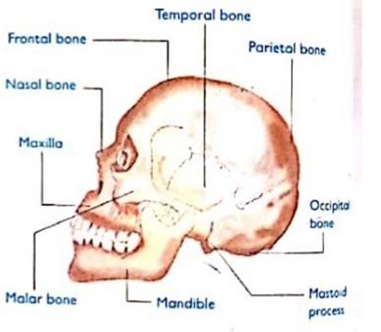
1. THE SKULL
Is the bony framework of the head.
The skull is made of cranial bones and facial bones
8 Cranial bones joined by immovable joints called sutures to form cranium
Cranium houses (covers) the brain, middle ear and part of the ear
Facial bones make up the upper jaw (maxilla) and lower jaws (mandible)
FUNCTIONS OF THE SKULL
(i) It protects the brain, olfactory organs, the eyes and the middle and inner ear.
(ii) It gives shape of the head.
THE DIAGRAM OF HUMAN SKULL
2. THE RIBS
Are thin, flat curved bones that form a protective cage around the organs in upper body.
Ribs comprise 24 bones which are arranged in 12 pairs.
The union between the ribs, vertebral column and the sternum makes the ribcage
Functions of the ribs
i. To give the chest its shape.
ii. To protect the heart, lungs, spleen and kidney against injuries and shock.
iii. Helps in breathing by expanding to let air in and contract to let air out.
Adaptations of the ribs
i. Ribs have long shaft for attachment of intercostal muscles
ii. Ribs have tuberculum and capitulars for articulation with tubercular and capitulars facets of the thoracic vertebrae.
iii. Ribs have curved shaft to provide a long surface area for attachment of intercostal muscles.
iv. Ribs have hard shaft for support and protection of delicate organs of thoracic cavity from mechanical damage.
TYPES OF RIBS
Ribs are of three categories namely;-
i. True ribs
ii. False ribs
iii. Floating ribs
TRUE RIBS
These are the first 7 pairs of ribs, at the back they are connected to the backbone.
At the front they are connected to the breast bone or the sternum.
FALSE RIBS
These are the next 3 pair of ribs.
They are slightly shorter than the true ribs.
At the back they are connected to the backbone, in the front they are not connected to the sternum, instead they are connected to the lowest rib.
FLOATING RIBS
These are the last 2 pairs of ribs.
They are the smallest of all.
They are attached to the backbone at the back but are not attached to anything in the front, hence the name floating ribs.
NB: The first seven ribs are attached directly to the sternum ventrally while the next three ribs are joined together ventrally to form costal cartilage which is then attached to the sternum.
The sternum is composed of small bone known as sterna brae
3. VERTEBRAL COLUMN
Is a series of 33 bones called vertebrae.
Vertebral column is also called the spine, backbone or spinal column.
Functions of vertebral column
i. Protects the spinal cord
ii. Supports the body trunk
DIAGRAM OF VERTEBRAL COLUMN
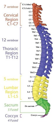
TYPES OF VERTEBRAE
Vertebral column has five types of vertebrae, namely:
i. The cervical vertebrae
ii. Thoracic vertebrae
iii. Lumbar vertebrae
iv. Sacral vertebrae
v. Caudal vertebrae
vi. Each vertebra is separated by an intervertebral disc of cartilage.
Function of intervertebral disc
i. Prevents wearing out of vertebrae during locomotion.
ii. Acts as a shock absorber
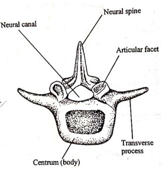
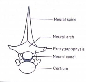
STRUCTURE OF THE VERTEBRAE
Each vertebra has the following parts:
| PARTS | DESCRIPTION | FUNCTION |
| 1. CENTRUM | Is the main body of the vertebra | Holds and supports other parts of the vertebra |
| 2. NEURAL CANAL | Is the hollow part just above the centrum | Allows passage of the spinal cord |
| 3. NEURAL ARCH | Is the part surrounding the neural canal | Protects the spinal cord Projects to form processes for muscles attachment. |
| 4. TRANSVERSE PROCESSES | These are lateral projections on the neural canal | Offers surface area for muscles attachment |
| 5. NEURAL SPINE | Projection from the back of the neural arch | Offers surface area for muscles attachment |
| 6. ARTICULAR FACETS | These are articulating surfaces include prezygapophysis and postzygapophysis | Offers articulation surface with adjacent bones and vertebrae |
DIAGRAM OF THE BASIC STRUCTURE OF VERTEBRA
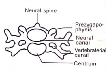
THE CERVICAL VERTEBRAE
These are short bones found in the neck region à They are seven in man.
The first two cervical vertebrae are known as ATLAS and AXIS
FUNCTION OF ATLAS AND AXIS
They permit movement of the head.
For example
The joint between the atlas and skull allows up and down (nodding) movements of the head
The joints between the atlas and axis allow turning or sideways movements of the head.
DIFFERENCES BETWEEN ATLAS AND AXIS
| ATLAS | AXIS |
| (i) Has no centrum | Has a wide centrum |
| (ii) Has no odontoid process | Has the odontoid process (projection of centrum) |
DIAGRAM OF CERVICAL VERTEBRAE
Functions of cervical vertebrae
(i) They provide the site for neck muscles to attach
(ii) They support the skull or weight of the head.
(iii) They allow free rotation/nodding of the skull on vertebral column
Adaptations of cervical vertebrae
i. They have vertebraterial canal for passage of vertebral artery and vertebral nerves.
ii. They have wing-like transverse processes for neck muscles attachment.
iii. They have short neural spine for attachment of neck muscles.
iv. They have large and wide neural canal for passage of spinal cord.
v. The atlas has facets that articulate with the skull to allow nodding movement.
vi. Axis has odontoid process to permit turning of the head
THE THORACIC VERTEBRAE
These are found in thorax region articulating with ribs.
They are 12 in man and 13 in rats.
DIAGRAM OF THORACIC VERTEBRAE
Function of thoracic vertebrae
They provide the site for muscles in the thorax to attach
Adaptations of thoracic vertebrae
i. They have long neural spine which offers a large surface area for the attachment of back muscles.
ii. They have short transverse processes for articulation with the ribs.
iii. They have prominent centrum to support the weight of the vertebrae above them.
iv. They have wide neural canal for passage of spinal cord.
v. They have facets for articulation with ribs
vi. They have articular surfaces (pre- and post-zygapophysis) covered with cartilage which is found between adjacent vertebrae. Cartilage reduces friction between adjacent bones.
THE LUMBAR VERTEBRAE
Are vertebrae between the lower end of the rib and waist.
Lumbar vertebrae found in the lumbar region of the body.
Most of mammals have 7 but man has 5 lumbar vertebrae.
Distinctive features of lumbar vertebrae
i. They have short and broad neural spine
ii. They have long transverse processes e.g. Extra transverse processes.
iii. They have large and enlarged centrum with a D-shaped neural canal.
Function of lumbar vertebrae
i. Provides site for abdominal muscles to attach
ii. Permits bending, sideways movements and rotation of the trunk
DIAGRAM OF LUMBAR VERTEBRAE
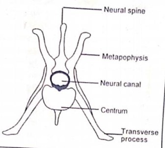
Adaptations of lumbar vertebrae
i. They have short and broad neural spine for attachment of powerful back muscles.
ii. They have long, large and well developed transverse processes for abdominal muscles to attach.
iii. They have projections (metapophysis and anapophysis) for increasing the surface area for muscles to attach.
iv. They have large and thick centrum for supporting the upper body weight.
THE SACRAL VERTEBRAE
These are situated in the sacral region (between the waist and tail).
They are three in most mammals but are 5 in man, all fused together to form sacrum.
DIAGRAM OF SACRAL VERTEBRAE

Distinctive features of sacral vertebrae
i. They have short neural spine.
ii. The sacrum is broader on the front side and narrow towards the tail.
iii. They have a small neural canal.
iv. The transverse processes of first sacral vertebrae are large and wing- like for articulation with pelvic girdle.
v. They have pairs of holes.
Adaptations of sacral vertebrae
Anterior vertebrae have a well- developed transverse process which are fused to the pelvis girdle or articulate with pelvic girdle.
i. The sacral vertebrae are fused for strength and transmit weight of the stationary animal to the rest of the body.
ii. Sacrum has broad base and short spine for attachment of back muscles.
THE CAUDAL (COCCYGEAL) VERTEBRAE
Caudal vertebrae are found in the tail region.
Their numbers differ from animal to animal depending on the animal’s size of the tail.
Since man has no external tail there are only four caudal vertebrae which are fused together to form the coccyx known as vestigial tail.
Distinctive features of caudal vertebrae
i. They have reduced transverse processes
ii. They have reduced neural spines and zygapophysis.
iii. They lack a neural arch.
THE APPENDICULAR SKELETON
The appendicular skeleton comprises the upper extremity and the lower extremity.
The upper extremity consists of following parts:
— Forelimbs (arms)
— Pectoral girdle
The lower extremity consists of the following parts:
— Hind limbs(legs)
— Pelvic girdle
NB: All mammals have limbs which are designed in the same plan of pentadactyl limb plan.
Pentadactyl limb means each limb ends with five digits (fingers or toes).
DIAGRAM TO SHOW PENTADACTYL LIMB PLAN

1. FORELIMBS (arms)
These are attached to the axial skeleton to the anterior part of the body. Forlimbs comprises the following parts
i. Humerus
ii. Radius and ulna
iii. Carpals, metacarpals and phalanges
DIAGRAM TO SHOW THE FORE LIMB
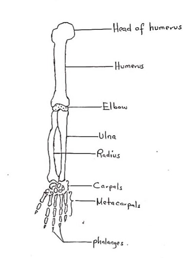
THE HUMERUS
Is a single long bone found in the upper arm.
It lies between the shoulder and the elbow
FUNCTIONS OF HUMERUS
Used for attachment of biceps and triceps muscles.
ADAPTATIONS OF HUMERUS
i. Has a round head that articulates with the glenoid cavity of the scapula forming a ball and socket joint.
ii. Has two rough projections, the greater and lesser taberosities near the head for muscles attachment.
iii. Has depression called bicipital groove between the two tuberosities for muscle attachment
iv. Has trochlea with a deep groove that fits in the sigmoid notch of ulna to form a hinge joint at elbow.
RADIUS AND ULNA
These are bone found in the forearm and are usually fused in rabbits.
Radius is on the side of the thumb while the ulna is on the side of the small finger.
Ulna has a projection called olecranon process, which has a sigmoid notch for articulation with humerus.
FUNCTION OF RADIUS AND ULNA
i. They support the carpals, metacarpals and phalanges.
ii. They provide surface area for attachment of muscles of the arm.
CARPALS, METACARPALS AND PHALANGES
Carpals
Are small bones which form the wrist
They articulate with radius and ulna at the upper and metacarpal at the lower end.They are 8 in man an 9 in rabbit
Metacarpals
Are small bones which found in the palm à They are longer than the carpals.
They are 5 in man, 3 in each finger and 2 in the thumb
Phalanges
These are the finger bones.
They are 14 in man
2. THE PECTORAL GIRDLE
This is also called the shoulder girdle
It is made up of two separate halves
Each half is made up of three bones that are attached to the vertebral column by ligaments and muscles.
Three bones of the pectoral girdle are:
1. Scapula
2. Coracoids
3. Clavicles
THE SCAPULA
Is a large triangular flat bone on the side of the rib cage.
It is also known as a shoulder blade.
The scapula has a concave depression called the glenoid cavity
Glenoid cavity articulates with the head of humerus to form a ball and socket joint.
FUNCTIONS OF THE SCAPULA
Provide site for attachment of muscles that move the arms.
It also connects the arm to the axial skeleton.
DIAGRAM OF SCAPULA
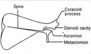
ADAPTATIONS OF THE SCAPULA
i. They have the glenoid cavity, a socket which articulates with the humerus forming a ball and socket joint at the shoulder.
ii. They have acromium and metacromium for muscles attachment
iii. The socket (glenoid cavity) has cartilage. These are the smooth surfaces to reduce friction.
iv. They are broad and flattened to increase the surface area for muscles attachment.
v. They are hard to provide support.
CORACOID
Is the bone of the pectoral girdle that is found at the apical end of the scapula à This bone is formed by coracoids process of the scapula.
THE CLAVICLES
These are also called the collar bone.
It is slender and s-shaped.
It connects the upper arm to the trunk of the body and has a shoulder joints.
They articulate anteriorly with the sternum and posteriorly with the acromion processes of the scapulate.
FUNCTIONS OF THE CLAVICLES
i. They provides site for muscles attachment.
ii. They also aid in movement of the arm.
THE PELVIC (HIP) GIRDLE
This consists of two hip bones known as pubic bones
Pubic bones fused ventrally to form pubis symphysis.
Parts of pubic bones
Each pubic bone comprises of three bones:
i. Pubis
ii. Ischium
iii. Ilium
DIAGRAM OF PELVIC GIRDLE
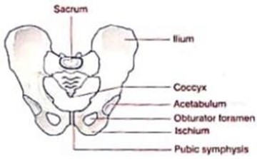
NB: These bones are completely fused together in adult.
The pelvis has a socket called the acetabulum which articulates with the femur to form the hip joint
The size of pubic cavity is very important in females during birth
During birth a hormone known as relaxin cause relaxation of the pubis symphysis thus expanding the size of the pelvic cavity.
FUNCTIONS OF PELVIC GIRDLE
i. It supports the weight of the body from the vertebral column
ii. It support and protects organs in the lower body such as the urinary bladder and the reproductive organs.
iii. It protects the developing foetus in a pregnant woman.
iv. Provides a large surface area for attachment of muscles that move the leg.
THE HIND LIMBS
These are attached to the axial skeleton to the posterior part of the body.
Hind limbs comprises of the following parts:
i. Femur (thigh bone)
ii. Tibia and fibula
iii. Tarsal, metatarsals and phalanges
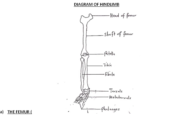
Is the longest, largest and strongest bone found in the thigh between the hip and the knee.
On the upper end, femur has a round head which fits into the acetabulum of the pelvic girdle to form a ball and socket joint of the hip.
The lower end of femur possesses two curved convex surfaces called condyles for articulation with tibia and fibula to form a hinge joint at the knee.
FUNCTIONS OF FEMUR
i. The femur supports the upper part of the body.
ii. Its shaft provides surface for attachment of thigh muscles.
KNEE CAP
This is a large, triangular bone between the femur and tibia.
Its main function is to protect the knee joint.
TIBIA AND FIBULA
These are bones that form the skeleton of the lower hind limb.
They are found between the knee and the ankle.
The fibula is fused to the tibia on the lower part of the leg
| TIBIA | FIBULA |
| (i) It is large and bears most of weight | It is small and serves as an area for muscles attachment |
| (ii) It is found in front (ventral) It is found on the side of the big toe | It is found behind (dorsal) |
ADAPTATION OF TIBIA AND FIBULA
i. Tibia has two slight depressions at the upper end, for articulation with the condyles of the femur.
ii. They have medial and lateral malleolus on the posterior end for articulation with tarsals bones.
iii. They are hard and strong shaft to support the body weight.
A MALLEOLUS
There are two types of malleolus namely:
i. Medial malleolus
ii. Lateral malleolus
MEDIAL MALLEOLUS
Is the prominence on the inner side of the ankle, formed by the lower end of the tibia.
LATERAL MALLEOLUS
Is the prominence on the outer side of ankle, formed by the lower end of the fibula.
DIAGRAM OF TIBIA AND FIBULA

TARSALS, METATARSALS AND PHALANGES
Tarsals
Are small bones which articulate with tibia and fibula to form the ankle joint
Metacarpals
Are small bones which articulate with metatarsals to form the foot.
Phalanges
Are small bones which articulate with metatarsals to form toes.
NB: Metatarsal and phalanges are similar in number and position to those of the hand.
ADAPTATIONS OF THE HUMAN SKELETON FOR CARRYING OUT ITS
FUNCTION
| Function | Adaptation |
| Support | à Firm and strong framework which maintains the shape of the body |
| Protection | à Presence of endoskeleton which is hard and firm. E.g. skull protects the organ in the head |
| Locomotion | à Presence of joints and muscles. |
| Synthesis of red and white blood cells | à Presence of bone marrows |
Questions: Explain how skeleton is adapted for the following functions.
i. Locomotion
ii. Synthesis of blood cells
iii. Protection
JOINT
is the point where two or more bones meet.
Joints are very important for movement and locomotion as they allow freedom of movement between bones
TYPES OF JOINTS
There are three types of joints namely;-
(i) Immovable/fixed joints
(ii) Gliding /slightly movable joints
(iii) Free movable/synovial joints
IMMOVABLE JOINTS
These are joints that do not allow any movement.
Immovable joints are also called fixed joints
They have no cartilage in them but bones are held closely together by short connective tissue fibres.
Example of immovable joints
Sutures the joints that found in the skull (cranium joints)
The joints between the bones of the
GLINDING JOINTS
These are joints that allow little movement.
Gliding joints are also called slightly movable joints or sliding bones
In this type of joint, short bones glide or slide over each other to bring about movement
Gliding joints have no fluid between them instead they have a large cartilage between them known as intervertebral disc which reduces friction during movement
Examples of gliding joints
Joints that occur between the vertebrae.
Joints found at the wrist and ankle
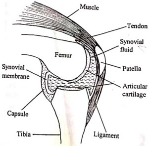
FREELY MOVABLE JOINTS
These are joints that allow a large degree of movement.
Freely movable joints are also called synovial joints
They have a slippery fluid secreted by synovial membrane called a synovial fluid
Function of synovial fluid
i. It reduces friction by lubricating the bones
ii. Acts as a shock absorber during movement.
TYPES OF MOVABLE JOINTS
There are three types of synovial joints, namely:
i. Ball and socket joints
ii. Hinge joints
iii. Pivot joints
BALL AND SOCKET JOINTS
These types of joint allow movement in all directions.
These joints involve two bones, one with a rounded head and the other with a depression into which the head of the first bone fits and move freely.
Examples of ball and socket joints
Hip joint ( a joint found between the femur and the pelvic girdle)
Shoulder joint ( a joint found between the humerus and the pectoral girdle)
DIAGRAM OF BALL AND SOCKET JOINT

HINGE JOINTS
These are the joints that allow movement in one direction only à Hinge joints look like the door. Examples of hinge joints
Elbow joint
Knee joint
DIAGRAM OF HINGE JOINT
PIVOT JOINT
Is the joint which allows movement in several directions.
In this type of joint one bone form a peg that enters into a cavity in the other bone.
The peg acts as a pivot over which the other bone rotates.
Example of pivot joint
Joint found between the atlas and the axis of the cervical vertebrae.
NB: Odontoid process of the axis fits into the neural canal of the atlas to form a pivot joint which allows rotational movements of the head.
MUSCLES
Muscle is a contractile tissue specialized for contraction and relaxation to bring about movement in the body.
Muscles tissue covers the skeleton.
Muscles are responsible for locomotion and any other type of movement in animals.
A muscle is a specialized tissue consisting of sheets of cells referred to as muscle fibres.
Muscles are capable of contraction so as to produce about movement or tension in the body.
TYPES OF MUSCLES
In the human body there are three types of muscle. These are;-
i. Cardiac muscles
ii. Smooth muscles
iii. Skeletal muscles
CARDIAC MUSCLES
These are muscles that are found only in the heart.
Their cells have a single nucleus
These muscles are controlled by involuntary nervous system.
Question
Why the heart beats continuously throughout the life of an organism?
Answer: Due to the presence of cardiac muscles that contract without suffering from fatigue
DIAGRAM OF CARDIAC MUSCLES
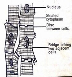
APTATIONS OF CARDIAC MUSCLES
i. They have abundant mitochondria to provide adequate ATP for contraction.
ii. They are myogenic to contract and relax without nervous stimulation.
iii. They are branched and interconnected to provide a large surface area for contraction and relaxations.
iv. They are multinucleated for better coordination of its contractile activities.
v. They are striated to allow for contraction and relaxation in short intense bursts
vi. They are elastic to allow for contractions and relaxation.
They have intercalated disc to allow the cardiac potential to travel across them, making it easier for electric impulses to move quickly.
Intercalated disc also acts as shock absorber to protect the myocorolium from mechanical shock.
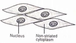
SMOOTH MUSCLES
These are the muscles found in the walls of organs such as alimentary canal, the blood vessels and the bladder
Smooth muscles also reffered to as involuntary muscles. This is because their activity is not under the control of the will.
They are made up of cells which are long and spindle-shaped
The cells have one central nucleus
DIAGRAM OF SMOOTH MUSCLES
ADAPTATIONS OF SMOOTH MUSCLES
i. They are connected by autonomic nervous system (involuntary nervous system) hence involved in involuntary actions.
ii. They have numerous mitochondria to provide ATP energy for contractions.
iii. They have spindle –shaped cells to allow smooth uniform contractions.
iv. They have elastic myofibrils to allow for contraction and relaxation.
SKELETAL MUSCLES
These are muscles attached to skeleton
Skeletal muscles are also known as voluntary muscles
Skeletal muscles are responsible for locomotion and other voluntary movement of the body.
ADAPTATIONS OF SKELETAL MUSCLES
i. They are multi-nucleated to allow for better control of contractile activities.
ii. They are long to offer a large surface for contraction and relaxation.
iii. They are striated to allow contractions and relaxations on short intense bursts for efficient locomotion.
DIAGRAM OF SKELETAL MUSCLE
MUSCLES AND MOVEMENT
Skeleton on its own cannot bring locomotion or any movement without the help of muscles.
GROUPS OF MUSCLES IN HINDLIMBS AND FORELIMBS
The hind limb has seven groups of muscles which are responsible for a wide range of movement of the legs. These are:
i. Protractor muscles
ii. Retractor muscles
iii. Adductor muscles
iv. Abductor muscles
v. Rotator muscles
vi. Flexor muscles
vii. Extensor muscles
I. Protractor muscle
These muscles pull the base of the leg forward.
II. Retractor muscles
These muscles pull the base of the leg backwards
III. Adductor muscles
These muscles pull the leg inwards towards the body.
Adductor muscles are also known as depressor muscles
IV. Abductor muscles
These muscles pull the leg outwards away from the body.
Abductor muscles are also known as levators
V. Rotator muscles
These muscles are responsible for rotating either the whole leg or a part of the leg at a joint.
VI. Flexor muscles
These muscles pull two parts of the leg towards each other.
VII. Extensor muscles
These muscles pull two parts of the leg away from each other.
The forelimb has two groups of muscles which bring about movement. These are:
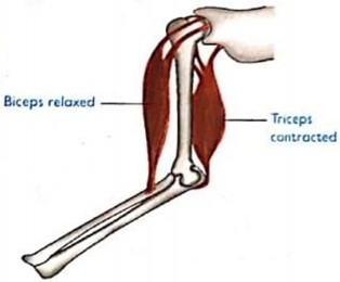
(i) Biceps
(ii) Triceps
Biceps
These muscles are also known as flexor muscles
Biceps is responsible for bending of the arm by pulling the radius and ulna upward.
Triceps
These muscles are also known as extensor muscles
Triceps is responsible for strengthening of the arm by pulling two limb bones away from each other.
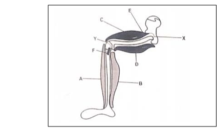
HOW MUSCLES FACILITATING MOVEMENT
Muscles work in an antagonistic fashion means they work oppositely to each other.
When one muscle contracts the other muscle relaxes
For example: When the biceps muscles contract, triceps muscles relax, radius and ulna are moved upwards, causing the arm to bend.
Biceps and triceps are known as antagonistic muscles because they work oppositely to each other.
ROLES OF MUSCLES IN MOVEMENT OF ARM
i. Strengthening of the arm (downward movement)
ii. The triceps muscles contract hence shortening à Biceps muscles relax (extends).
iii. Radius and ulna moves downwards, and the arm straightened.
DIAGRAM SHOWING MUSCLES DURING
STRENGTHENING OF THE ARM
Bending of the arm (upward movement)
Biceps muscles contract
Triceps muscles relax
Radius and ulna are moved upwards, causing the arm to bend.

GENERAL ADAPTATIONS OF THE MUSCLES
i. They are very elastic for stretching or contracting of muscles
ii. They are supplied with blood vessels for nutrients supply and carrying wastes away from the muscles.
iii. Their cells produce ATP energy for the muscles to function
MUSCLE CRAMPS
Muscle cramp is an involuntary contraction of the skeletal muscles.
Muscle cramp is often painful and can last from seconds to 10 minutes.
Muscle cramp may involve part of a muscle or an entire muscle.
CAUSES OF MUSCLE CRAMPS
The following are some common causes of muscle cramps:
i. Vigorous activity
The vigorous use of a muscle in sports or in any other physical activity may cause muscle cramps during the activity or even hours later.
ii. Injury
A muscle cramp may occur as a protective mechanism following an injury such as broken bone. This occurs to minimize movement and stabilize the area of injury.
iii. Dehydration
à When one loses more body fluids and salts, mostly water than the amount that is taken in, muscle crap may occur.
iv. Muscle fatigue
When the muscle contract powerfully may result muscles fatigue very quickly hence muscle cramp may occur.
v. Lack of magnesium or calcium in the body
Magnesium and calcium are important in muscle contraction and therefore lack of them they lead to muscle cramps.
vi. Lack of oxygen in the muscles (inadequate of oxygen muscles).
EFFECTS OF MUSCLE CRAMPS
i. Muscle cramps cause a lot of pain.
ii. Leads to tenderness and firmness of the involved muscles
iii. May disrupt the functioning of the involved body organ. For example when a muscle cramp occurs in the foot it can cause difficult in walking.
PREVENTION OF MUSCLE CRAMPS
Muscle cramps may be prevented through;-
1. Stretching the affected muscle more often. E.g. standing up and walking around can stop cramps in the leg.
2. Gently massaging the muscle so as to relax it.
3. Drinking a lot of water before, during and after the activity.
4. Taking of salt in solution form or licking to replace the amount of salt lost in the body.
5. Rapid breathing as well as stretching the muscles can improve cramps from lack of oxygen.
6. To do a lot of physical exercise before engaging in sports or other physical activity.
7. Exercise activity especially during warm weather should be avoided.
9. Applying warmth from a warm cloth could also be a quick remedy.
MOVEMENT IN PLANTS
Movement in most plants is very slow thus unnoticeable.
Most plant movements are growth movements and these movements are referred to as growth curvature or movement of curvature.
Movement of curvature
Is movement that involves certain parts of the plant while the plant is fixed on its position
Parts of the plant that show movement
i. Leaves
ii. Stems
iii. Roots
NB: These parts they move when they grow and enable plants to obtain their requirements such as water and light.
QuestionExplain why plants do not need to locomote as animals?
Answer: Plants do not need to locomote as animal because:-
i. They obtain water and nutrients from the soil through their roots.
ii. They are capable of manufacturing their own food through photosynthesis.
iii. Most perennial plants can survive harsh conditions such as drought by shedding their leaves or having deep roots to absorb water in deeper soils
iv. Fertilization is aided by pollination through wind insects.
TYPES OF GROWTH MOVEMENT IN PLANTS
Growth movement in plants is divided into two categories, namely:-
(i) Autonomic movement
(ii) Paratonic movement
I. AUTONOMIC MOVEMENT
These are self-controlled growth movements. Example of automic movement
Growth in the meristematic region, i.e. tips of shoots and roots.
II. PARATONIC MOVEMENT
These are the plant growth movements induced by external stimuli such as light, moisture, gravity, temperature, touch and chemicals.
TYPES OF PARATONIC MOVEMENT
Paratonic movement is divided into two categories, namely:
i. Tropic movement
ii. Nastic movement
I. TROPIC MOVEMENTS
These are the plant growth movements in response to the stimuli.
Tropic movement is also called tropism.
Tropism is the directional growth of a plant organ in response to an external stimulus such as light.
The plant moves either towards or away from the stimulus.
— If the movement is towards the stimulus, it is called positive tropism.
— If the movement is away from the stimulus, it is called negative tropism.
TYPES OF TROPIC MOVEMENTS
The following are types of tropic movement
(i) Phototropism
(ii) Hydrotropism
(iii)Geotropism
(iv) Thigmotropism
(v) Chemotropism
i. Phototropism is the plant growth movement in response to the source of light.
ii. Hydrotropism is the plant growth movement in response to the source of water.
iii. Thigmotropism is the plant growth movement in response to the source of touch.
iv. Chemotropism is the plant growth movement in response to the source of chemicals.
v. Geotropism is the plant growth movement in response of the source of gravity.
vi. Thermotropism is the plant growth movement in response to the source of heat.
II. NASTIC MOVEMENT
These are non-directional movement of plant organs in response to diffuse stamens are independent of external stimuli
Example of nastic movement
i. Folding of leaves in warm weather conditions
ii. Opening and closing of flowers in response to intensity of light.
iii. Closing of leaves when attached.