COORDINATION
Coordination is the working together of different parts of the body in an orderly and organized manner
Without coordination the body becomes disorderly and it may fail to function properly.
IMPORTANCE OF COORDINATION
1. Coordination ensures survival of organisms.
2. Coordination enables organism to detect their life necessities such as food for heterotrophs, detection of light by autotrophs.
3. Coordination helps living organism to respond to their stimuli.
IRRITABILITY OR SENSITIVITY
Is the ability to perceive, interpret and respond to changes in the internal and external environment.
1. External environment
This is outside surrounding of whole organisms.
Components of external environment
The following are components of external environment:
i. Light
ii. Sound
iii. Pressure
iv. Gravity
v. Chemicals
vi. Water
vii. Food
2. Internal environment
This is the surrounding at cells within the body of an organism.
Components of internal environment
The following are components of internal environment:
i. Water
ii. Glucose
iii. Minerals
iv. Ions
v. pH
vi. Temperature.
COMPONENTS OF COORDINATION
There are five components of coordination namely:
i. Stimulus
ii. Receptors
iii. Coordinators
iv. Effectors
v. Response
I. STIMULUS ( plural: Stimuli)
Is the change in the environment of an organism
TYPES OF STIMULI
There are two types of stimuli, namely:-
a. External stimulus
b. Internal stimulus
A. EXTERNAL STIMULUS
Is the stimulus which is associated with the surrounding environment
Example of external stimuli
> Heat
> Wind
> Pressure
> Chemicals
> Water
> Food
> Light
B. INTERNAL STIMULUS
Is the stimulus which occurs within the organism
Example of internal stimuli
> Water
> Glucose
> Mineral ions
> pH
> Temperature
II. RECEPTORS
Are the specialized cells that detect stimulus
In animals receptors are located in specialized organs known as sense organs.
Example of receptors
> Receptor for pain, touch, heat, and cold-are located in the skin
> Receptors for taste-located in the tongue
> Receptors for light-located in the eye
> Receptors for sound-located in the ear
> Receptors for smell-located in the nose
— When a receptor detects stimuli, it creates impulses which are transmitted to the coordinating system through nerve cells.
NERVE IMPULSE
Is a slight electric charge which travels along a nerve cell
III. A COORDINATOR
Is an organ that receives messages from the receptors, translates them and sends the information back to effectors for action.
Example of coordinators
1. The brain
2. Spinal cord
IV. EFFECTORS
Are the parts of the body that respond to the stimuli.
Example of Effectors
1. Muscles
2. Glands
3. Cilia
4. Flagella
V. RESPONSE
Is a behavioural, physiological or muscular activity initiated by a stimulus
OR
Is the change shown by an organism in reaction to a stimulus
Examples of response
— Blinking when an insect lands on the eye
— Dropping a hot object.
The table below shows the relationship between some stimuli, receptor, effectors and response
| Stimuli | Receptors | Effectors | Responses |
| Heat | Skin | Skin | Secretion of sweat, sweating |
| Cold | Skin | Skeletal muscles | Uncontrolled contraction and relaxation of skeletal muscles, shivering |
| Skin | Formation of goose pimples. | ||
| Taste | Tongue | Salivary glands | Secretion of saliva, salivation |
| Pain | Skin | Skeletal muscles | Contract, move organs away from source of pain |
| Sound | Ear | Ear drum | Hearing of noise, music or sound. |
The sequential order of transmissions of a nerve impulse from a sensory organ to the organism’s response is
![]()
The Ways in Which Coordination is Brought About
Coordination is controlled or effected by two major systems, namely:
i. Nervous system
ii. Hormonal system called endocrine system
> The coordination in simple multi-cellular animals is controlled by nervous system only.
> The coordination in higher animals called vertebrates (including human beings) is controlled by nervous system and endocrine system.
> Coordination in plants is under the control of hormones.
NERVOUS COORDINATION IN HUMAN, NEURONES
NEURONES: Are cells which carry electrical impulses from the central nervous system to all parts of the body
Neurone is the basic unit of the nervous system.
Neurones are also called nerve cells
STRUCTURE AND FUNCTIONS OF THE NEURONE
Each neurone consists three basic features, namely;-
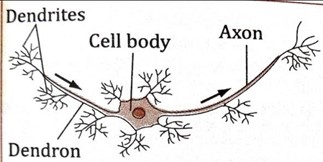
i. The cell body
ii. Dendrites
iii. The axon
Other parts/features of the neurone are:
(i) Dendrons
(ii) Myelin sheath
(iii) Schwann cells
(iv) Node of Ranvier
(v) Axoplasm

CELL BODY
Is the main part of the nerve cell
Function of the cell body
i. It gives rise to other parts of the nerve cell
ii. It is the main control centre of the nerve cell
Components of the cell body
The cell body has the following components:
(i) Cytoplasm– enclosing the nucleus.
(ii) Nucleus– which control all activities
(iii) Mitochondria– that provide energy for metabolic processes.
DENDRITES
Are short numerous fibres which receive nerve impulses from other neurones and transmit them to the cell body.
THE AXON
Is the elongated fibre that extends from the cell body The longer the axon, the faster it transmits information.
The role of axon
It transmits nerve impulses away from the cell body.
MYELIN SHEATH
Is a fatty layer that covers axon for protection and insulation
Function of myelin sheath
i. Protects the neuron and allow impulses to travel faster.
ii. It insulates the axon
SCHWANN CELLS
Are cells found on the surface of myelin sheath
Function of Schwann cells
i. They secrete the myelin sheath
NODES OF RANVIER
Are constrictions which interrupt myelin sheath at exactly one millimeter interval
Role/function of node of ranvier
i. Used to speed up the transmission of impulses.
DENDRONS
Are extensions of the cell body
They form branches known as dendrites
Function of Dendrons
i. They transmit impulses towards the cell body
AXOPLASM
Is a specialized type of cytoplasm which is continuous with the cytoplasm in the cell body
Function of axoplasm
i. It is a part through which nerve impulses travels
NEURILEMMA
Is a layer of cells which encloses the myelin sheath
ADAPTATION OF NEURONES TO THEIR FUNCTION
1. They have numerous mitochondria for energy supply during conduction of impulses
2. They are long so as to enables transmission of impulses to long distance in the body.
3. They have node of Ranvier to increase the speed of impulse transmission
4. They are supplied with denser network of blood capillaries for supply of food and oxygen
5. They are numerous for effective transmission of impulses along the whole body.
6. They are covered with fatty myelin sheath for protection and insulation.
7. They have numerous dendrites for connectivity with other neurons.
8. They have Schwann cells that secrete myelin sheath.
9. They have elongated axons which help in quick transmission of impulses.
NB: The axon terminates into synaptic knobs.
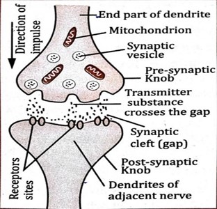
These knobs have vesicles containing a chemical transmitter substance, for example acetylcholine.
The axon of one neuron and the dendrites of the next neuron do not actually touch each other.
The gap between neurons is called the synapse
A SYNAPSE
Is a junction between two neurones
Function of synapse
i. It enables impulse to be passed from one neurone to another.
ii. It ensures that impulses are transmitted in one direction only.
THE TRANSMISSION OF NERVOUS IMPULSES ACROSS A SYNAPSE
The transmission of nervous impulses across a synapse is mediated by chemical substances called neurotransmitter
Example of neurotransmitters
i. Acetylcholine
ii. Noradrenaline
The transmission of nervous impulses across synapses occurs as follows:
i. When an impulse reaches the synaptic knob of the pre-synaptic neurone, synaptic vesicles discharge the neurotransmitter into the synaptic cleft.
ii. Where it diffuses across the cleft and binds to specific receptors on the post-synaptic membrane.
iii. This leads to generation of action potential in the post-synaptic membrane.
iv. The result is the transmission of an impulse along the post-synaptic neurone.
QUESTION: Why the nerve impulse travels only in one direction?
REASON: This is because the neurotransmitters are found only on the pre-synaptic knob meaning that impulses can only travel from the pre-synaptic neuron to the post-synaptic neuron.
TYPES OF NEURONES
There are three types of neurons, namely:
1. Sensory neurons
2. Motor neurons
3. Relay (intermediate) neurons
Each of these neurons has a different structure and performs different functions.
1. SENSORY NEURONES
Are nerve cells that transmit impulses from the sensory receptors to the central nervous system.
Sensory neurones have their cell bodies off the axon and outside the central nervous system.
Sensory neurons are also called afferent neurones
Function of sensory neurons
i. They transmit impulses from a receptors to the central nervous system

TYPES OF SENSORY NEURONES
There are two types of sensory neurons, namely:
i. Visceral sensory neurones: are those neurones that transmit nerve impulses from internal organs
ii. Somatic sensory neurones: are those neurones that transmit impulses from the skin, skeletal muscles, joints and bones
2. RELAY NEURONES
Are nerve cells that connect sensory neurone and motor neurone in the central nervous system
Relay neurons are located in the central nervous system between the sensory and the motor neurons.
Relay neurones are also called intermediate neurones
Function of relay neurones
To convey messages between neurones in the central nervous system.
DIAGRAM OF RELAY NEURONE
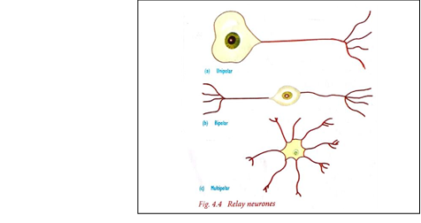
TYPES OF RELAY NEURONES
i. A unipolar neurone: is a type of neurone that has its axon extending from its cell body.
ii. A bipolar neurone: is a type of neurone that has an axon and dendrons extending in two different directions from the cell body.
iii. A multi-polar neurone: has one axon and several dendrons extending from the cell body in different directions.
NB: The axon extends to the motor neuron
3. MOTOR NEURONES
Are nerve cells that transmit impulses from the central nervous system to the effectors
The cell body of a motor neurone is at one end of the neurone and lies entirely within the central nervous system.
It has tiny branches at each end (dendrites) and a long fibre (axon) that carries the signals or nervous impulses.
Motor neurones are also called efferent neurones
Function of motor neurones
i. To transmit impulses from the central nervous system to the effectors.

STRUCTURAL DIFFERENCES BETWEEN MOTOR AND SENSORY NEURONES
MOTOR NEURONE | SENSORY NEURONE |
| (i) The cell body is located at one end of the neurone | The cell body located near one end of the neurone |
| (ii) It is multipolar | It is unipolar |
| (iii)Has a large and irregular cell body | Has small and definite cell body |
| (iv) Has dendrites surrounding cell body | Has no dendrites which surround cell body |
NERVOUS SYSTEM
This system is made up of the brain, spinal cord and nerves.
Parts of the nervous system
Nervous system is divided into two parts, namely:
1. Central nervous system
2. Peripheral nervous system
1. CENTRAL NERVOUS SYSTEM (CNS)
Is the part of the nervous system consisting of the brain and spinal cord.
It coordinates all the neural functions.
THE COMPONENTS OF THE CENTRAL NERVOUS SYSTEM AND THEIR FUNCTIONS
The central nervous system has two main components, namely:
i. The brain
ii. Spinal cord
THE BRAIN
Is a delicate organ enclosed within a body structure called the skull or cranium.
Brain is the master control of the body.
Brain is covered by a system of membrane called meninges.
The brain is the main centre for integrating and coordinating impulses.
Function of human brain
i. The human brain is a specialized organ that is ultimately responsible for all thought and movement that the body produces.
ii. It allows humans to successfully interact with their environment, by communicating with others and interacting with inanimate objects near their surroundings. For example If the brain is not functioning properly, the ability to move, generate accurate sensory information or speak and understand language can be damaged as well.
PARTS OF THE HUMAN BRAIN
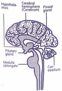
The human brain is divided into three parts, namely:
1. Fore brain
2. Mid brain
3. Hind brain
DIAGRAM OF HUMAN BRAIN
1. FORE BRAIN
Is the anterior portion of the brain
The outer portion is grey hence called grey matter and inner portion is whitish hence called white matter.
Fore brain is responsible for voluntary actions
Fore brain is made up of:
i. Cerebrum.
ii. Hypothalamus
iii. Thalamus
iv. Pituitary gland
v. Olfactory lobes
I. CEREBRUM
Is the largest part of the human brain
Cerebrum is covered by a thin layer of grey matter called cerebral cortex
Functions of the cerebrum
The cerebrum has the following functions:
i. It is responsible for reasoning and intelligence.
ii. It is involved in learning, imagination and creativity.
iii. It is the memory centre.
iv. It is responsible for personality or character.
v. It controls voluntary body movement such as walking and dancing.
vi. It is responsible for sight, hearing, taste, smell and speech.
Parts of the cerebrum
Cerebrum is divided into two parts (cerebral hemispheres), namely:
i. Right hemisphere
ii. Left hemisphere
Right hemisphere
Is the part of the cerebrum which sends and receives impulses from the left side of the body
Left hemisphere
Is the part of the cerebrum which sends and receives impulses from the right side of the body
HYPOTHALAMUS
This part is concerned with body temperature and osmoregulation.
> It contains osmoreceptors and thermoreceptors that detect changes in osmotic pressure and internal body temperature respectively.
> It has a very rich blood supply
Function of the hypothalamus
i. It coordinates and controls the autonomic nervous system
ii. It has centers that control appetite, thirst and sleep.
iii. It also controls the activities of pituitary gland.
iv. It acts as an endocrine gland.
PITUITARY GLAND
This is the master of endocrine glands.
Function of pituitary gland
i. It secretes hormones which control osmoregulation, growth, metabolism and sexual development.
OLFACTORY LOBES
Is the part of fore brain that receives impulses of smell via olfactory nerves from the nose.
Function of olfactory lobes
i. It is concerned with the sense of smell.
2. MID BRAIN
Is the smallest part of the brain which found between the fore brain and hind brain.
The mid brain consists of the optic lobes, which are the main area for audio and visual processing.
Functions of the midbrain
i. To relay information between the fore brain and hind brain.
ii. To relay information between fore brain and the eye through optic nerves.
iii. It is responsible for the movement of the head and trunk.
THALAMUS
The thalamus is located in the middle part of the brain.
i. It helps to control the attention span, sensing pain.
ii. It monitors input that moves in and out of the brain to keep track of the sensations the body is feeling.
iii. It contains the centre for the integration of sensory information.
3. HIND BRAIN
It is made up of cerebellum and the medulla oblongata
CEREBELLUM
Is located in front of medulla oblongata
The cerebellum controls essential body functions such as balance, posture and coordination, allowing humans to move properly and maintain their posture.
Functions of the cerebellum
i. It maintains posture, movement and balance
ii. It ensures that all muscles work together to produce smooth coordinated voluntary movement.
iii. It assists in the learning of new motor skills like playing the piano, swimming and riding a bicycle.
NB: Damage to the cerebellum results in uncoordinated movements
MEDULLA OBLONGATA
This is the central part of the autonomic nervous system
Function of medulla oblongata
i. It controls all unconscious activities of the body e.g. Breathing, heartbeat, digestion, dilation and contraction of blood vessels, secretion of juices from glands and temperature regulation
ii. It contains a number of reflex centre for regulating heartbeat, breathing, blood pressure.
iii. It controls swallowing, salivation, vomiting, coughing, and sneezing.
SPINAL CORD
Is a part of the central nervous system and is posterior to the brain.
The spinal cord extends from the base of the brain into the inside of vertebral column.
It is enclosed in meninges and protected by the vertebral column
THE STRUCTURE OF THE SPINAL CORD
Structurally the spinal cord consists of the following parts:
(i) Grey matter
(ii) White matter
(iii) Dorsal root
(iv) Ventral root
I. GREY MATTER
Is the central part of the spinal cord
It surrounds the central canal which is filled with cerebrospinal fluid
Grey matter consists of relay neurones which relay information between the afferent and efferent neurones.
The grey matter has cell bodies, dendrites and synapses
CEREBROSPINAL FLUID (CFS)
Is a clear watery fluid secreted from the blood
It is similar to lymph whose normal contents include glucose, salts, enzymes, and some white blood cells (but no red blood cells).
Function of cerebrospinal fluid
1. Provides nourishment to brain tissues
2. Serves as a shock absorber
II. WHITE MATTER
Is the outer part of the spinal cord surrounding the grey matter.
White matter consists of axon of sensory and motor neurones.
VENTRAL ROOT
This is a part of the spinal which carries motor nerve fibres.
III. DORSAL ROOT
This is a part of the spinal which carries sensory nerve fibres.
THE STRUCTURE OF SPINAL CORD

FUNCTIONS OF THE SPINAL CORD
i. Conducts sensory nerve impulses from the receptors to the sense organs to the brain
ii. Conducts motor nerve impulses from the brain to the effectors
iii. Enables animals to attain an upright posture through the maintenance of muscle tone
iv. It serves as the reflex centre for all spinal reflexes.
PERIPHERAL NERVOUS SYSTEM (PNS)
Is a system which is made up of a network of nerves linking various parts of the body to the brain and spinal cord
> It links the CNS with the receptors and effectors
THE COMPONENTS OF THE PERIPHERAL NERVOUS SYSTEM AND THEIR
FUNCTIONS
The peripheral nervous system (PNS) is divided into two parts (components):
a. The somatic nervous system
b. The autonomic nervous system
(a) THE AUTONOMIC NERVOUS SYSTEM
This comprises of nerves that control the involuntary activities of the body.
The autonomic nervous system is also called involuntary nervous system
The autonomic nervous system has sensory and motor neurons running between the central nervous system and various internal organs, for example the heart and lungs.
Example of involuntary activities of the body
> gut movements
> beating of the heart
> secretion of glands
Function of the autonomic nervous system
> It is responsible for involuntary control of internal organs, blood vessels and smooth muscles.
The autonomic nervous system is divided into:
i. Sympathetic nervous system.
ii. Parasympathetic nervous system.
THE SYMPATHETIC NERVOUS SYSTEM
This system normally produces effects that prepare the animal for emergency.
It activates the fight or flight’ response under sudden or stressful circumstances.
Example of effects produced due to sympathetic stimulation
i. Increasing the heart rate
ii. Dilating the pupils
iii. Increasing blood pressure
iv. Glucose formation in the liver
THE PARASYMPATHETIC NERVOUS SYSTEM
This system normally produces effects that aimed at energy conservation.
It helps the body to return to normal activity after an emergency
Example of effects produced during parasympathetic stimulation
i. Decreasing the heart rate
ii. Constricting the pupils
iii. Decreasing blood pressure
iv. Stimulation of the digestive tract
NB: Together, these two systems regulate homeostasis within the body – one preparing the body for action, and the other repairing the body afterward.
(b) THE SOMATIC NERVOUS SYSTEM
This comprises of nerves that control voluntary activities of the body.
The somatic nervous system is also called voluntary nervous system
The somatic system has neurones connected with voluntary skeletal muscles and sense organs.
Example of somatic nervous system
— Nodding of the head
— Raising the leg
Function of the somatic nervous system
i. It is responsible for the voluntary control of the skeletal muscles, bones and sense organs.
ii. It is composed of afferent nerves that carry information to the central nervous system (spinal cord) and efferent nerves that carry neural impulses away from the central nervous system.
Peripheral nervous system consists of the following nerves:
i. Cranial nerves
ii. Spinal nerves
CRANIAL NERVES
These nerves arise from the brain and link the brain to the structures in the head, neck and upper of the trunk.
i. There are 12 pairs of cranial nerves.
Function of cranial nerves
i. They transmit impulses to the sense organ and muscles of the head and neck.
Examples of cranial nerves
— The optic
— Auditory
— Olfactory nerves.
SPINAL NERVES
These arise from the spinal cord.
They are associated with receptors and effectors in the trunk for example the arms and legs.
In humans, there are a total of 31 pairs of spinal nerves.
Each spinal nerve has two roots namely: dorsal root and ventral root
REFLEX ACTION
Is a rapid involuntary response to a certain stimulus
Reflex actions are rapid and happen without us thinking.
Example of reflex action
The withdrawal of the hand from a hot or sharp object Sneezing in response to a foreign body in the nose Blinking of the eye incase an object passes close to it.
i. Salivation
ii. Swallowing
iii. Secretion of tears when an onion is cut
iv. Enlargement of pupil in different light intensities
THE NEURONIC PATHWAY (REFLEX ARC) OF A REFLEX ACTION
REFLEX ARC: Is the route that is followed by impulses during a reflex action
OR
Is the path taken by impulses involved in the reflex action
Components of a reflex arc (neuronic pathway)
A reflex arc consists of the following components;
i. Sensory receptor
ii. Sensory neurone
iii. Relay neurones
iv. Motor neurone
v. Effectors e.g. muscle or gland
NB: Relay neurone is within the central nervous system (CNS)

Sequence of a reflex action
The receptors in the sense organs are stimulated by the stimulus and sensory nerve impulse is generated. The impulse is transmitted through a sensory nerve fibre to the central nervous system.
The nerve impulse is received by the intermediate neurones within the central nervous system (CNS)
The motor nerve impulse is transmitted to the effectors through a motor nerve fibre from the central nervous system.
Then the effectors respond.
TYPES OF REFLEX ACTION
Basically there are two types of reflex action, namely
(i) Simple reflex
(ii) Conditioned reflex.
I. SIMPLE REFLEX ACTION
Is a quick involuntary response to stimulus.
It occurs without conscious thought and does not need to learn.
Example of simple reflex actions
i. Blinking
ii. Salivation
iii. Coughing
iv. knee jerk
v. Sneezing
vi. Dropping a hot object.
QUESTION: What will happen when a person touches a hot object?
ANSWER: When a person touches a hot object, simple reflex action occurs without conscious thought as follows:
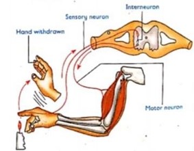
i. The heat from a hot object stimulates pain receptors in the skin. This generates an impulse which is transmitted by the sensory neurone to the spinal cord.
ii. In spinal cord, the impulse is integrated and interpreted then the impulse is transmitted to the motor neurone via the relay neurone.
iii. The impulse travels along the motor neurone to the biceps of the arm, which contracts resulting in withdrawal of the hand from the hot object.
The diagram below shows a simplified reflex arc of this situation
CONDITIONED REFLEX ACTION
Is a learned response resulting from a stimulus from a past experience which was originally ineffective in producing a specific response
This response develops over a period of time.
Example of conditioned reflex action
Seeing a banana may not have any effect on someone who has never seen a banana before. But after one has eaten several bananas and found them to be sweet, seeing a banana will cause salivation.
The conditioned reflex was first described experimentally by Ivan Pavlov a Russian scientist using dogs as follows:
i. He observed the sight or smell of food initiates salivation in dogs. This is a normal reflex called the salivation reflex
ii. He rang a bell whenever he was feeding his dogs. He continued doing this for several weeks. Later on, he rang the bell in absence of food. He found that this stimulated salivation in the dogs. Thus, the original stimulus (sight or smell of food) was replaced by a different and unrelated stimulus (ringing the bell) through learning.
iii. Conditioned reflexes are mediated by the brain through previous experience.
iv. In case of Pavlov’s experiment, the dogs had learnt to associate ringing of the bell with the presence of food.
v Therefore, ringing the bell initiated the same response as the presence of food.
vi. Conditioned reflexes can also be unlernt. If ringing of the bell is continued but this time in absence of food, dogs will stop salivating on hearing it.
DIFFERENCE BETWEEN SIMPLE REFLEX AND CONDITIONED REFLEX ACTION
| SIMPLE REFLEX ACTION | CONDITIONED REFLEX ACTION |
| (i) It is inborn response to external stimuli | It is a learnt response |
| (ii) Does not involve the brain directly | It involves the brain directly |
| (iii)It is the same in all members of a species | It differs among members of a species |
| (iv) Single stimulus brings about response | Combined stimuli (related and unrelated stimuli) brings about response |
| (v) Initiated by a related stimuli | Initiated by an unrelated stimuli |
| (vi) It is always constant | It can be reinforced through rewards and punishment |
SENSE ORGANS
Sense organ is a complex specialized organ or structure where sensory neurones are concentrated and function as a receptor to specific stimuli.
TYPES OF SENSE ORGANS
In mammals, there are five main sense organs, namely:-
(i) The eye – for sight
(ii) The ear – for hearing
(iii) The nose – for smell
(iv) The skin – for pressure, touch, temperature and pain
(v) The tongue – for tasting
THE SENSORY RECEPTORS
These are specialized cells that detect stimulus
Types of sensory receptor
According to their location, there are two types of sensory receptors, namely:
i. Interreceptor
ii. Exteroceptors
INTERORECEPTORS
Are sensory receptors which are located within the body They respond to stimulus from within the body.
Example of interoreceptor
— Osmoreceptors
EXTERORECEPTORS
Are receptors which are located near the body surface
> They respond to stimulus from the external environment
Example of exteroreceptor
— Mechanoreceptors
TYPES OF RECEPTORS
According to the stimulus which responds to, there are several types of receptors, namely:
(i) Photoreceptors
(ii) Thermoreceptors
(iii) Chemoreceptors
(iv) Pain receptors
(v) Mechanoreceptors
(vi) Osmoreceptors
i. Photoreceptors:
are cells sensitive to light
ii. Mechanoreceptors: are cells sensitive to pressure and vibration
iii. Thermo receptors: are cells sensitive to temperature
iv. Chemoreceptors: are cells sensitive to chemical substance
v. Pain receptors: are cells sensitive to pain on the surface and in the body.
vi. Osmoreceptors: are cells sensitive to osmotic pressure
1. THE TONGUE
Is an organ responsible for taste
It has a specialized group of sensory cells called taste buds.
Test buds are also called gustatory cells
In mammals, taste buds occur on raised portions of the upper surface of the tongue called lingual papillae while in other vertebrates, they are distributed on the walls of buccal cavity.
They have hair-like processes projecting above the surface of the papillae.
The tongue is kept moist by saliva, and is richly supplied with nerves and blood vessels.
Role of test buds
Helps in taste detection of the food taken.
MAIN TASTES THAT THE HUMAN TONGUE IS ABLE TO DETECT
There are four main tastes that the human tongue is able to detect. These are:
Sweet: detected at the tip of the tongue.
Sour: detected at the sides of the tongue.
Bitter: detected at the back of the tongue.
Salt: detected all over the tongue.

ADAPTATIONS OF THE TONGUE TO ITS FUNCTIONS
The tongue is adapted to its functions by possessing the following features:
1. The tongue has taste buds which help it to respond the stimuli such as sweet, bitter, sour and salt.
2. At the base of each taste bud there is a nerve that sends the sensations to the brain.
MECHANISM OF FOOD TASTE DETECT IN THE TONGUE
1. When the food is in the mouth, taste buds become stimulated by the chemicals dissolve on the moist surface of the tongue and therefore generate a nerve impulse.
2. The nerve impulse generated is taken to the brain via sensory fibres and produce sensation of taste such as sweet.
NB:
i. The tongue is always moist to dissolve the incoming chemicals from food for sensation of taste.
ii. The sensory fibres are found at the base of the taste buds and connect the taste buds to the brain
ii. The combined activity of taste buds and smell receptors gives the sensation of flavor.
Importance of sensation of taste
i. It helps animals to distinguish between suitable from unsuitable substances for ingestion
ii. It stimulates the salivary glands to secrete saliva containing enzymes and stomach walls to secrete the gastric juice containing digestive enzymes.
iii. It helps one to enjoy good food and to reject bad food.
2. THE HUMAN NOSE
The nose: is an organ responsible for the sense of smell.
> It has a specialized group of sensory cells called olfactory cells located in the upper part of the nasal cavity.
> Nasal cavity is an air filled space found inside the nose Olfactory cells are cells sensitive to smell
> They are also called olfactory bulb
> The olfactory cells secrete mucus that moistens the nostrils.
THE MECHANISM OF SMELLING OCURS AS FOLLOWS:
When a substance dissolves in moisture caused by mucus in the olfactory region, the olfactory cells are stimulated and an impulse is sent to the olfactory lobes of the brain via the olfactory nerve. The nerve impulse is interpreted as smell.
QUESTIONS
Question 1:
Why when we have the cold we lose sense of smell?
Answer: Because the olfactory surfaces become dry.
Question 2: Why hot food often has more taste than cold food?
Answer: This is because it activates more the taste buds and the smell receptors.
Question 3: Why we cannot taste foods smell when suffering from cold?
Answer: This is because the nasal passages are inflammed and coated with mucus. The smell receptors are essentially non- functional.
ADAPTATIONS OF THE NOSE IN CARRYING OUT ITS FUNCTIONS
The nose able to carry out its functions due to the following features
i. It is made up of cartilage to allow flexibility of the nose during blowing or sneezing thus removing dust, particulate matter or microorganisms.
ii. It has sinuses containing mucous secreting cells for mucus secretion
iii. It has mucus produced by mucous secreting cells which keeps the inner surface of the nose moist and traps unwanted substances.
iv. It has olfactory nerve which sends impulses to the brain enabling us to sense smell.
The major functional differences between the taste receptors and smell receptors
Smell receptors are cells specialized for detecting vapour coming to the organism from distant source while taste receptors are cells specialized for detection of chemical present in the mouth.
Smell receptors are much more sensitive than taste receptors while taste receptors are much less sensitive than smell receptors
3. THE HUMAN SKIN
Is the largest organ in the body
As a sense organ, the skin senses touch, pressure, pain, heat and cold.
It protects us from microbes and the elements
Helps to regulate body temperature and permits the sensations of touch, heat, and cold.
THE STRUCTURE OF THE SKIN
Structurally, the mammalian skin is made of two main layers, namely:
1. Epidermis
2. Dermis
1. THE EPIDERMIS
Is the outer layer of the skin which contains melanin
Melanin is a pigment which determines the colour of the skin and protects the body against ultra violet radiations.
It contains dead cells that protect the body against bacterial invasion and reduces loss of water through evaporation.
The epidermis is made up of three layers, namely:
i. Cornified layer
ii. Granular layer
iii. Malpighian layer
I. CORNIFIED LAYER
Is the outermost layer of the epidemis
It is made up of keratinised dead cells that prevent entry of bacteria, physical damage and loss of water through evaporation.
II. GRANULAR LAYER
Is the middle layer of the epidermis.
It is made up of living cells that give rise to the cornified layer.
III. MALPIGHIAN LAYER
Is the innermost part of the epidermis
It is made up of actively dividing cells that give rise to new epidermal cells.
It contains melanin pigment.
Function of melanin pigment
i. Determines the colour of the skin
ii. Protects the inner layers of the skin against ultra violet radiations.
2. THE DERMIS
Is the inner layer of the skin
It is made up of collagen fibres and elastic fibres which gives the skin toughness and flexibility and fat cells which store energy and provide thermal insulation.
It is comparatively thicker than the epidermis.
The dermis contains the following structure:
a. Sweat glands
b. Blood capillaries
c. Nerve ending
d. Sensory cells.
SWEAT GLANDS
These are coiled tubules which have long ducts opening to the surface through the pores.
Function of sweat glands
They secrete sweat through pores in the skin surface.
The way on how sweat glands produce sweat
The secretory cells in sweat glands absorb excess water, mineral salt, traces of urea, lactic acid and carbon dioxide from the blood capillaries, and then secrete them as sweat into the surface of the skin.
A. BLOOD CAPILLARIES
These are blood vessels which supply the skin with nutrients and oxygen and remove excretory products.
B. NERVE ENDINGS
These consist of sensory nerve cells that detect changes from the external environment.
They are sensitive to touch, pressure, cold, heat and pain.
C. HAIR FOLLICLES
These are lined with granular and malpighian layers of the epidermis.
The hair follicles hold the hair fibre
They are supplied with sensory nerves to increase sensitivity of the skin.
The base of each hair follicle is connected to the epidermis by an erector Pilli muscles which contract to make hair stand and relax to make hair lie.
D. SEBACEOUS GLANDS
Are glands that produce an oily chemical substance called sebum
They are attached to the hair follicles and drain their contents into the hair follicle.
Function of sebum
i. Acts as antiseptic to bacteria therefore, it protects the skin against disease causing microorganisms (pathogens).
ii. It keeps the hair and epidermis supple, flexible and water proof.
E. SUBCUTANEOUS LAYER
This is a fat layer that lies beneath the dermis.
It is also known as adipose tissue.
Function of subcutaneous layer
i. It acts as heat insulating layer
ii. It provides a channel for blood vessels and nerves to the skin.
iii. It acts as a storage region for fats and food reserves.
THE STRUCTURE OF THE SKIN

FUNCTIONS OF SKIN
i. Protects the underlying tissues from physical damage and prevents the entry of microorganisms.
ii. It helps in body temperature regulation
iii. It helps in excretion of excess water mineral salts and traces of urea through sweat
iv. It synthesizes vitamin D through the action of sunlight. Ergosterol in the fatty layer of the skin converts into vitamin D under the influence of sunlight.
v. Responds to external stimuli such as heat, cold, pain and touch.
vi. Prevents excessive loss or absorption of water since it is water proof.
vii. Produces melanin pigment that protects the body from ultra violet radiations.
viii. The skin acts as sensory organ due to the presence of various nerve endings.
ix. It prevents micro-organism and other foreign materials from entering the body.
x. It acts as a storage organ for fats in the body.
ADAPTATIONS OF THE SKIN TO ITS FUNCTIONS
1. It has cornified layer made up of dead cells to prevent entry of bacteria, physical damage and desiccation.
2. It has granular layer made up of living cells that give rise to cornified layer.
3. It has melanin pigment in the malphigian layer which protects the body against ultra violet
4. It has blood vessels in the dermis which supply oxygen and nutrients to tissues of the skin and remove excretory products
5. It has sebaceous glands to produce sebum which is antiseptic to bacteria
6. It has the hair erector muscles which controls whether the hair stands erect or lies down depending on the temperature of the surrounding.
7. It is supplied with nerves which convey impulses to the central nervous system to be interpreted.
8. It has blood vessels in the dermis which dilate when the body temperature is high to facilitate heat loss by radiation and constrict when the temperature is low to reduce heat loss.
9. It has sweat glands to produce sweat which helps to cool down the body.
10. It has sensory cells which are sensitive to touch, pressure, cold enable response to environmental changes.
11. It has subcutaneous fat or adipose tissue which acts as heat insulating layer.
12. It has sweat glands to secrete sweat which contain water, sodium chloride, uric acid and urea through pores in the skin surfaces hence acts as an excretory organ.
SKIN AS A SENSE ORGAN
Skin as a sense organ is composed of the following sensory receptors:
NB: The skin contains sensory nerve endings which are receptors.
They are sensitive to pain, pressure, touch, heat and coldness.
When the nerve endings are stimulated they set up nervous impulses which are sent to the spinal cord or brain to be interpreted.
i. Touch receptors
ii. Pressure receptors
iii Pain receptors
iv. High temperature receptors
v. Cold receptors
Skin receptors are more complex consisting of nerve endings called encapsulated nerve endings surrounded by a connective tissue.
Function of the connective tissue
i. It protects the nerve ending from mechanical damage
ii. It helps in generation of a nerve impulse.
The encapsulated nerve endings include:
i. Meissner’s corpuscles – respond to touch
ii. Krausser’s end organs – respond to cold
iv. Ruffini’s end organ – respond to pain
v. Pacinian corpuscles – respond to pressure
vi. End bulls corpuscles – respond to heat
Below is a table summarizes the functions and locations of receptors in the skin
| RECEPTOR | LOCATION | FUNCTION |
| Touch receptors | On the superficial part of the skin | Sensitive to touch |
| Pressure receptors (Pacinian corpuscles) | In the lower part of the dermis | Sensitive to pressure |
| Pain receptors | Free nerve ending below the epidermis | Sensitive to pain thus protects the body against potential harm by initiating reactions such as reflex action. |
| High temperature recptors | In the middle of the dermis | Sensitive to temperatures between 30C to 43C |
| Cold receptors | At the base of dermis | Sensitive to temperatures between 20C to 30C |
NECTA PRACTICAL 2A (2020) question
1. You are provided with a tooth pick, piece of cotton wool, methylated spirit and samples labelled A and B which are stimuli of receptors in your body. Carry out the experiments in term (i) – (iv) and then answer the questions that follow:
Sample A– Sands
Sample B– wheat flour
i. Look at your body and observe the sense organ that covers the whole hands.
ii. Take a tooth pick and prick slightly the upper part of your hand and note the feeling.
iii. Touch each of the samples A and B and feel their coarseness
iv. Take cotton wall and soak into methylated spirit. Rub it on your hand and observe what is happening.
4. THE MAMMALIAN EYE
Is a complex light sensitive organ specialized for sight
It is spherical in shape
It contains numerous light sensitive cells called photoreceptors in a specialized region known as retina
The eyeball is located in a cavity in the skull called orbit or eye socket.
FUNCTION OF ORBIT
i. It offers protection of eyeball against physical damage.
ii. It has thick layer of fat deposited around the eyeball, which serves as a shock absorber.
iii. The eyeball is attached to the walls of the socket by a pair of antagonistic muscles that control its movement.
MUSCLES THAT CONTROL MOVEMENT OF EYEBALL
There are two pairs of antagonistic muscles that control movement of eyeball, namely:
i. Superior and inferior oblique muscles – moves the eyeball left and right.
ii. Superior and inferior rectus muscles – moves the eyeball up and down.
iii. These muscles can move the eyeball in many directions to increase the field of view.
PARTS ASSOCIATED WITH THE EYE
a) EYELIDS
These are two thin folds of the skin found in front of the eyeball.
The movement of the eyelids is known as blinking.
Blinking keeps the surface of the eye moist.
The eyelids have tear glands or lacrimal glands
There are two types of eyelids namely
i. Upper eyelids
ii. Lower eyelids
Function of eyelids
They protect the external surface of the eye.
TEAR GLANDS
They are found below the upper eyelid of each eye Function of tear glands
They secrete a saline liquid (tears)
A tear is a solution of sodium chloride and hydrogen carbonate.
Tears contain enzymes which kill microorganisms and protect the eyeball from infection.
Tears drain into the nose through small tubes at the corner of the eye called canaliculi into the pharynx.
Blinking washes this liquid across the surface of the eyeball.
Function of tears
i. To keep the surface of the eyeball moist.
ii. To prevent the eyeball from friction.
iii. To protect the eyeball from infection.
EYE LASHES
These are relatively many long hairs found on the edge of the eyelids.
Function of eye lashes
They protect the eyeball from foreign particles such as dust and insects.
EYEBROWS
These are hairs above the eyelids that prevent sweat from the forehead and dust from entering the eye.
Function of eyebrows
They prevent the entry of dust particles and sweat into the eye.
STRUCTURE OF THE EYE
The eye is made up of the following parts
1. Sclera
2. Cornea
3. Conjunctiva
4. Choroid
5. Ciliary body
6. Iris
7. Pupil
8. Lens
9. Aqueous humour
10. Vitreous humour
11. Suspensory ligaments
12. Retina
13. Optic nerve
14. Blind sport

FUNCTIONS AND ADAPTATION OF PARTS OF THE EYE
SCLERA: Is the outermost layer of the eye
Sclerotic layer is white in colour and is composed of elastic connective tissues.
It is the tough opaque layer of the eye
It is made of dense collagen fibres.
Function of the sclera
i. It protects the eye
ii. It supports and maintains the shape of the eyeball.
NB: Sclera continues and becomes transparent layer at the front of the eye to form cornea.
CORNEA
Is the transparent front part of the sclera that allows light into the eye
Cornea is covered by a thin membrane known as conjunctiva.
Function of cornea
i. It is convex (curved) to refract light.
ii. It is transparent to allow light to pass through.
CONJUNCTIVA
Is a thin transparent membrane that covers cornea
It lies in the inner surface of the eyelids
Function of the conjunctiva
i. It is transparent to allow the light to enter the eye.
ii. It is tough to protect the inner parts of the eye from mechanical damage.
CHOROID
Is a layer in the eye between the sclera and the retina
It is heavily pigmented layer
Choroid extends to the front of the eye to form the ciliary body and iris.
Function of the choroid
i. It has a dark pigment which prevents internal reflection in the eye by absorbing scattered light ray
ii. It contains a dense network of blood vessels, which supply oxygen and nutrients to the eye and remove metabolic waste products.
IRIS
Is a ring of contractile muscles which is continuous with the ciliary muscles.
It is the coloured visible part of the eye
Iris has two sets of muscles namely:
i. Circular muscles
ii. Radial muscles
NB: Contraction and relaxation of the circular and radial muscles control the size of the pupil by dilation and constriction thus controlling the amount of light entering the eye
Function of the iris
(i) Controls the amount of light entering the eye.
(ii) Its pigment absorbs light to prevent blurred vision
(iii) Its pigment determines the colour of the eye.
CILIARY BODY
Is made of contractile muscles which control the shape and curvature of the lens so as to improve focusing.
The contraction and relaxation of the ciliary muscles changes the shape of the lens.
Functions of ciliary body
i. They have glandular cells which secretes the aqueous humour
ii. They control the shape and curvature of the lens
PUPIL
Is the hole in the centre of the iris through which light enters into the eye
Function of the pupil
(i) It allows light to enter the eye.
NB: In dim light, the pupil dilates to allow more light into the eye while in bright light the pupil constricts to allow less light into the eye.
LENS
Is a transparent biconvex elastic structure filled with a jelly like substance
It is attached to the ciliary body by suspensory ligaments.
Function of the lens
i. It is transparent to allow light to pass through.
ii. It is biconvex to refract light onto the retina.
iii. It is elastic in nature to allow it to change shape when the eye focusing on near and far objects.
iv. It separates the aqueous humour from the vitreous humour
AQUEOUS HUMOUR
Is the watery transparent fluid found between the cornea and the lens.
Function of aqueous humour
i. It refracts light onto the retina
ii. It is transparent to allow light to pass through
iv. It maintains the shape of the eyeball
iv. Oxygen and nutrients diffuse from the blood vessels to nourish the cornea and the lens
VITREOUS HUMOUR
Is a dense clear gel that fills the posterior chamber of the eye between the lens and retina.
Function of vitreous humour
i. It maintains the shape of the eyeball.
ii. It is transparent to allow light to get to the retina
SUSPENSORY LIGAMENTS
Are inelastic structures that hold the lens in position
Function of suspensory ligaments
i. They attach the lens to the ciliary muscles
ii. Hold the lens in position
RETINA
Is the innermost part of the eye which contains light sensitive cells called photoreceptor cells.
Function of retina
i. It is a layer of the eye where the image is formed.
ii. It is a part of the eye where light is detected
Types of photoreceptor cells found in the retina
Retina has two types of photoreceptor cells namely:
1. Cones
2. Rods
CONES
Are cells sensitive to light of high intensity
Cones contain a photochemical pigment (light-sensitive pigment) called iodopsin
Iodopsin is adapted for bright light vision and colour vision.
Cones are able to detect colour
Cones are densely packed together in a certain region of the retina known as the fovea or yellow spot.
RODS
Are cells sensitive to light of low intensity
Rods contain a photochemical pigment known as rhodopsin
Rhodopsin is adapted for dim light vision and does not detect colour
Rods are able to detect black and white colours only.
Rods are scattered all over the retina but are more numerous on the periphery. Because of this, one can see an object better in dim light if he looks at it from the corner of the eye.
DIFFERENCES BETWEEN CONES AND RODS
| CONES | RODS |
| (i) They are sensitive to light of high intensity | They are sensitive to light of low intensity |
| (ii) They have pigment known as iodopsin | They have pigment known as rhodopsin |
| (iii)They are used for colour vision | They are used for night vision |
| (iv) Found in fovea | Found in other parts of retina. Not found in fovea |
QUESTION: Explain why nocturnal animals are able to see properly during night than during the day?
ANSWER: Because they have large number of rods in their retina which enable them o see clearly during night.
FOVEA
Is a small depression on retina that has cones only
Fovea is also called a yellow spot
Function of fovea
It a region for colour detection
BLIND SPOT
Is an area in the retina through which optic nerve leaves the eyeball.
Blind spot has neither rods nor cones, so image from objects falling on the blind spot cannot be perceived by the brain.
QUESTION
Why the blind spot is not sensitive to light?
ANSWER: Blind spot is not sensitive to light because it has no rods or cones.
Differences between the fovea and blind spot of the eye
| FOVEA | BLIND SPOT |
| (i) It has cones | It has no cones |
| (ii) It is sensitive light | It is not sensitive to light |
OPTIC NERVE
Is a cranial nerve which contains sensory neurons.
The neurons transmit impulses from rods and cones on the retina to the brain for interpretation
Optic nerve leaves the eye at the blind spot
ACCOMMODATION OF THE EYE
Accommodation
Is the ability of the eye to focus both near and distant objects.
It is a reflex mechanism which enables the eye to adjust and bring an image from a far or near object to focus on the retina.
Accommodation ensures that clear images of objects are formed.
MECHANISM OF ACCOMODATION
Accommodation of the eye is accomplished through a change in the shape of the lens
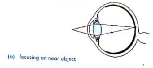
When the eye is focusing on a near object, the ciliary muscles contract while the suspensory ligaments relax, the lens becomes thick and allows light rays from near objects to be focused on the retina.
Consider the diagram below
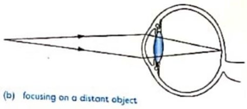
When the eye is focusing on a distant object, the ciliary muscles relax while the suspensory ligaments contract. The lens becomes thin, light rays from far object are less refracted and hence focused on the retina.
Consider the diagram below
RESPONSE TO LIGHT INTENSITY
The amount of light entering the eye is determined by the size of the pupil
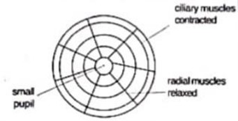
In bright light, the circular muscles of the iris contract while radial muscles relax reducing the size of pupil. This limits the amount of light entering the eye.
Consider the diagram below
In dim light, the radial muscles of the iris contract while circular muscles relax increasing the size of pupil thus allowing more light to enter the eye.
Consider the diagram below

BILOGICAL SIGNIFANCE OF THE ABOVE RESPONSE
(i) It protects the retina from damage by excessive light.
(ii) It improves visibility in dim light
Questions:
1. (a) Explain how the iris regulates amounts of light entering into the eye. Or explain how the iris of the eye responds to light of varying intensity.
(b) What is the purpose of the response you have described in (a) above
IMAGE FORMATION AND INTERPRETATION
An image is formed on the retina when the photoreceptors (rods and cones) are stimulated.
Mechanism of image formation
When light falls on an object, it is reflected. Some of the reflected rays fall on the eye.
The light from the object is refracted by cornea, aqueous humour, lens and vitreous humour, focusing it on the retina. The image formed on the retina is upside down and smaller than the object. The image on the retina stimulates the photoreceptor cells causing the generation of impulses in neurones. Impulses are then transmitted to the brain through the optic nerve for interpretation. A normal size, upright and coloured image is formed.
Consider the diagram below

ADAPTATIONS OF THE EYE
i. It has conjunctiva which protects the eyeball from mechanical damage and also helps the eyeball to move/ rotate easily by secreting mucus.
ii. It has cornea which is transparent to allow light into the eye and refracts the light entering the eye.
iii. It has aqueous and vitreous humours which are thin aqueous jelly- like fluids to allow light to pass through and refract it. They contain solutions of salts, sugar and proteins to provide nourishment to the eye.
iv. It has iris which is opaque and contractile for controlling the amount of light entering the eye by controlling the size of the pupil.
v. It has ciliary body contains ciliary muscles for controlling the shape and curvature of the lens
vi. It has suspensory ligaments to hold the lens in position.
vii. It has a transparent biconvex lens which allows light to pass through and to refract light onto the retina.
viii. It has retina which contains photoreceptor cells for image formation.
ix. It has rods which contain rhodopsin pigment for dim light vision.
x. It has cones which contain iodopsin pigment for colour vision, bright light and light of high intensity.
xi. It has fovea centralis with a high concentration of cones for accurate vision.
xii. It has the choroid layer to provide nourishment (oxygen and nutrients) to the eye.
xiii. Choroid also has black pigments to stop or reduce light reflection and absorb stray light.
xiv. It has a sclerotic layer to protect the inner more delicate parts of the eye and to give shape to the eye.
xv. It has optic nerves contain sensory neurones for transmission of impulses from retina to the brain for interpretation
xvi. It has pupil which is a gap between upper and lower iris through which light enters the eye.
xvii. An external eye muscle is contractile to move the eyeball (within the socket).
xviii. Eyelashes prevent dust and hazardous particles from reaching the conjunctiva.
COMMON DEFECTS OF THE MAMMALIAN EYE AND THEIR CORRECTION
Defects of the mammalian eye are structural deviations of the eye which alter the focusing mechanism of the eye.
There are two common eye defects, namely:
(i) Short-sightedness (myopia)
(ii) Long-sightedness (Hypermetropia)
I. SHORT-SIGHTEDNESS (MYOPIA)
Is the eye defect whereby a person cannot focus distant objects properly
A person is able to focus only near objects.
The myopia is caused by long eyeball which results the image to be formed in front of the retina.
Myopia can be corrected by using spectacles with concave lens (diverging lens).
Concave lenses diverge the light rays before they reach the eye.
Consider the diagram below

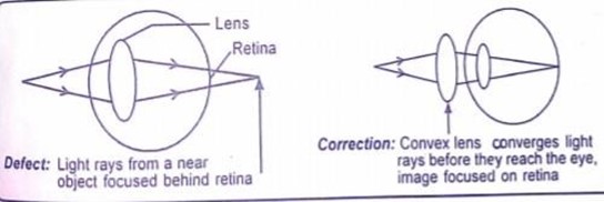
II. LONG-SIGHTEDNESS (HYPERMETROPIA)
Is an eye defect whereby a person cannot focus near objects properly.
A person is able to focus only distant objects.
Hypermetropia is caused by short eyeball which results the image to be formed behind the retina.
Hypermetropia can be corrected by using spectacles with convex lenses.
Convex lenses converge the light rays before they reach the eye.
Consider the diagram below
OLD SIGHTEDNESS (PRESBYOPIA)
Is a defect caused by loss of flexibility or elasticity of the lens and weakening of ciliary muscles.
Person cannot focus near objects properly.
The defects can be corrected by using spectacles with bifocal lenses.
ASTIGMATISM
Is a condition in which the cornea or lens is uneven such that images are not focused properly on the retina.
The defect can be corrected by using spectacles with special cylindrical lenses.
CATARACTS
Is a worm disease in which worms invade the lens
The lens becomes cloudy such that light cannot pass through easily and the person cannot see properly.
It is curable at early stage but if it is chronic lens may have to be removed by operation and can be replaced by a plastic lens inside the eye or a good eye from a donor.
CLAUCOMA
Is the eye defect caused by pressure in the eye
It is more common in old people
A person with this defect forms blurred images.
COLOUR BLINDNESS
Is the genetic disorder in which a certain colour cannot be distinguished by man.
A common type of colour blindness is red- green blindness, individual is not in position to determine/distinguish between red and green colour.
TRACHOMA
Is a viral disease which affects the lining of the eyelids.
Trachoma can be transmitted from one person to another through contact
If not treated, trachoma can cause blindness
5. THE HUMAN EAR
Is a specialized organ responsible for hearing and maintaining body balance.
The ear contains specialized sensory cells (receptors) that are sensitive to sound and the position of head with respect to gravity.
FUNCTION OF THE EAR
The mammalian ear performs two main functions
i. Hearing (detection of sound waves)
ii. Maintenance of body balance and posture
THE STRUCTURE OF THE EAR
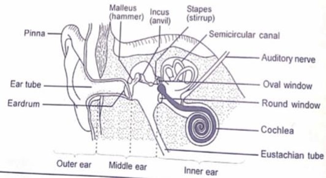
Structurally the ear is divided into three main parts (chambers), namely:-
i. The outer ear
ii. The middle ear
iii. The inner ear
Consider the diagram below showing the parts of ear
i. THE OUTER EAR
Is the external part of the ear filled with air
The outer ear is made up of the following parts
i. Pinna
ii. Auditory canal
iii. Eardrum
PINNA
Is the outermost part of the ear and is made up of cartilage.
It is a funnel-shaped flap
Function of pinna
a. It collects sound waves and directs them into the ear canal.
b. It helps some animals to determine the direction of sound. E.g. cattles
EXTERNAL AUDITORY CANAL (EAR CANAL)
Is a tube that directs sound waves to the eardrum.
Ear canal is also known as meatus
Functions of auditory canal
i. It is a tube through which sound waves travel.
ii. It has hairs and secretes wax, which help to trap dust and micro-organisms thus protecting the eardrum.
iii. It has sebaceous glands that secretes sebum in its lining to moisten the eardrum and lining of the canal
EARDRUM (TYMPANIC MEMBRANE )
Is a thin double membrane that forms the boundary between the outer and middle ears
It vibrates when hit by sound waves.
Functions of eardrum
It converts the sound waves into vibrations and transmits them to the ear ossicles.
2. THE MIDDLE EAR
Is an air-filled cavity in the skull.
The middle ear is made up of the following parts:
i. Ear ossicles
ii. Oval window
iii. Round window
iv. Eustachian tube
EAR OSSICLES
Are three small tiny bones that link the eardrum with the oval window.
They are named according to their shape
i. Malleus (hammer)
ii. Incus (anvil)
iii. Stapes (stirrup)
Function of ear ossicles
i. Used to amplify the vibrations
ii. Used to transmit amplified vibration of sound from the eardrum to the oval window
iii. Used to connect the eardrum to the oval window
OVAL WINDOW
Is a flexible membrane that covers a small hole leading to the inner ear.
Function of oval window
It transmits vibrations to the inner ear.
EUSTACHIAN TUBE
Is a narrow tube that connects the middle ear to the pharynx (mouth cavity).
Function of Eustachian tube
i. It allows air to get in and out of the middle ear
ii. It equalizes the air pressure between the middle and the outer ear hence preventing distortion (rupture) of the eardrum.
NB: The Eustachian tube is normally closed, but opens during swallowing, chewing and yawning.
3. THE INNER EAR
Is a fluid filled cavity consisting of a series of chambers and canals embedded in the bone of the skull.
The fluid in the inner ear is called perilymph
The following are chambers and canal in the inner ear
i. Semicircular canals, Utricle and Saccule
ii. Cochlea
SEMICIRCULAR CANALS
Are fluid filled tubular cavities and each has a swelling known as ampulla at one end.
Semicircular canals, utricle and saccule are also called vestibular apparatus
Function of semicircular canals, utricle and saccule
Help to maintain body balance and posture
COCHLEA
Is a coiled tube filled with liquid called endolymph
Cochlea contains sensory cells which are connected to the brain through the auditory nerves
The part of cochlea that responds to sound is called organ of corti
Function of cochlea
i. It is the structure responsible for sense of hearing.
ii. It is coiled to offer a large surface area for attachment of sensory cells responsible for hearing.
iii. It has sound receptors in the organ of corti to detect sound vibrations (hearing)
Function of organ of corti
It is a part of cochlea that responds to sound
MECHANISM OF HEARING
The pinna collects and directs sound waves into the auditory canal. From the auditory canal, the sound waves are passed on to the eardrum causing it to vibrate. The vibrations from the eardrum picked by malleus, incus and then to the stapes. The stapes passes the vibrations to the oval window which amplifies the sound waves 22 times. When the oval window vibrates, it causes the fluid in the inner ear and in the cochlea to move hence stimulates the sensory hair cells in the organ of corti. When the sensory hair cells become stimulated they generate nerve impulse. The impulse generated is transmitted to the brain via the auditory nerve. The brain interprets the impulse as sound of specific pitch and loudness.
Consider the diagram below

BALANCE AND POSTURE
The semi-circular canals, utricle and saccule are the structures concerned with animal’s sense of balance and position in space. The canals are filled with fluids which moves as the body moves or as the head changes position. The movement of the fluid stimulates sensory nerves in the canals and impulses are sent to the brain. The brain then sends impulses that affect an appropriate posture.
ADAPTATIONS OF THE MAMMALIAN EAR TO ITS FUNCTIONS
The ear is adapted to its functions by possessing the following features:
i. It has pinna which collects sound waves and directs them into the inner ear via the auditory canal.
ii. It has ear drum which converts the sound waves into vibrations and transmits them to the ear ossicles.
iii. The lining of auditory canal contains wax-secreting cells which produce wax to protect the inner delicate parts of an ear from mechanical damage.
iv. It has ear ossicles which amplify and transmit vibrations to the oval window.
v. It has Eustachian tube which allows air in and out of the middle ear to equalize the air pressure between the middle and the outer ear hence preventing rupturing of the eardrum.
vi. It has cochlea which is coiled to increase the surface area for sound reception.
vii. Presence of fluid-filled vestibular apparatus in the inner ear which facilitate balancing of sound when fluid is displaced.
DRUGS AND DRUG ABUSE IN RELATION TO NERVOUS COORDINATION
Drugs
are chemical substance which when taken into the body alter the structure or functioning of the body.
Examples of drugs
i. Cocaine
ii. Heroine
iii. Tobacco
iv. Marijuana
PSYCHOACTIVE DRUGS
Are the drugs that affect the central nervous system.
Psychoactive drugs produce a false sense of well-being and relieve someone from tension, anxiety, stress and pain.
TYPES OF PSYCHOACTIVE DRUGS
I. STIMULANTS
Are drugs which stimulate the nervous system.
They speed up brain activities and also the body processes.
Example of stimulants
— Cocaine
— Heroine
— Nicotine
— Caffeine from coffee, tea
II. SEDATIVES (DEPRESSANTS)
Are sleep- inducing drugs.
They slow brain activities and evoke sleep. E.g. alcohol,
III. PAINKILLERS
Are drugs immobilize or suppress pain centre in the brain
Painkillers are only prescribed by the doctors on inevitable cases because they cause brain damage.
IV. INHALANTS (VOLATILE SOLVENTS)
These include compounds like glue, kerosene, toluene and petroleum.
Other inhalants such as chloroform and industrial solvents are used as intoxicating drugs.
V. HALLUCINOGENS
Are drugs that distort the way the brain interprets impulses from the sensory organs.
These distortions may take one or two of the following forms
— The brain may alter the massage about something real, producing Illusion
— The brain may produce images with no basis in reality called hallucinations
Example of hallucinogens
Marijuana.
NB: Users of hallucinogens drugs may show signs of mental illness with confusion, violence and depression.
VI. NARCOTICS
These drugs dull the senses and relieve pain by depressing the cerebral cortex in the brain.
Narcotics also affect the thalamus, the body’s mood-regulating center. Example of narcotics
— Codeine
— Morphine
— Heroine
— Opium
FORMS OF DRUG TAKING
I. Intravenous: this is injecting a chemical substance into the blood system through a vein.
II. Inhalation: some people prefer to inhale volatile solvents such as petrol, glue or paint.
III. Oral: some other drugs like marijuana are smoked
IV. Sniffing: some drugs like cocaine are sniffed through the nose.
PROPER WAYS OF HANDLING AND USING DRUGS
1. Avoid taking any drug without diagnosing the disease and prescription by the doctor.
2. Always stay away from peer pressures and drug addicts to avoid copying their bad habits.
3. Keep yourself busy with a number of activities such as sports and games, reading books, etc.
4. Report any case of drug abuse or trafficking to concerned authorities.
5. Form a counselling club to advise people especially youths on how to keep off from drugs.
6. If one feels addicted, s/he should seek advice from health officials.
7. Never take a dose more or less that what has been prescribed by the doctor.
8. Complete the prescribed dose even after you start feeling well or after the symptoms of the disease has disappeared.9. Keep all drugs out of reach of children and drug addicts.
DRUG ABUSE
Is the misuse of drugs for the reasons other than the medical reasons.
OR is the use of a drug for any purpose other than what it was intended for.
When drugs are used regularly, they can cause a stable of dependence called addiction.
DRUG ADDICTION
Is a habitual and uncontrollable behaviour involving the use of drugs.
OR is a state of over dependence of drugs so that life becomes unbearable without it.
CAUSES OF DRUG ABUSE
i. Desire to satisfy curiosity about the effects of drugs.
ii. Sense of belongingness to a certain group.
iii. Desire to have a new life experiences.
iv. Some people just take drugs as an experiment to find out the experience the drug users feel, but badly end up becoming drug addicts.
v. To escape from life realities such as poverty, hunger, family quarrels.
vi. Peer pressure from peer groups.
vii. Peer pressure leads people to drug so as to create a sense of belonging and fitting in the peer group. It’s often said that teens use drugs when their friends do.
viii. Lack of education or ignorance
ix. Lack of employment
x. Lack of family upbringing
xi. For recreational purposes and excitements.
xii. Drug users believe that taking drugs make them feel better and lively.
xiii. To do away with unpleasant feelings and memories.
xiv. Some people take drugs as a way to forget problems and life hardships they experienced in life. Some people take drugs to avoid physical or emotional pain, discomfort, stress, boredom, anxiety and depression.
EFFECTS OF DRUG ABUSE
1. Excessive use of drugs can cause:
2. Health hazards
3. Social hazards
HEALTH EFFECTS OF DRUG ABUSE
1. Cigarette smoking can lead to lung cancer and other heart diseases
2. Bhang affects the reproductive system by slowing down the rate of sperm production
3. Alcohol causes brain damage, liver cancer and gastric diseases
4. Drugs e.g. miraa causes ulcers and rotten teeth
5. Many drugs affect the brain and give a false sense of happiness which is short lived.
6. Spread of diseases such as HIV/AIDS
7. Cocaine cause high blood pressure, heart failure and can lead death
8. Misuse of drugs weakens the body immune system
SOCIAL EFFECTS OF DRUG ABUSE
i. Students drop out of school
ii. Marriage breakdown
iii. Poverty
iv. Failure to concentrate at work
v. Increases of crime and violence
vi. Leads to irresponsible sexual behaviours
vii. Leads to accidents, loss of properties and death
PREVENTIVE AND CONTROL MEASURES OF DRUG ABUSE
1. Avoid taking any form of drug without prescription from the doctor
4. Enforcement of laws, rules and regulation for the control and supply of drugs.
5. Avoid emotional pain and stress by engaging in creative activities such as sports and games
6. The youth should be motivated to get involved in the fight against drug abuse.
7. Early detection, treatment and rehabilitation of drug addicts can help minimize the problem.
8. Various effects of drug addiction must be advertised through newspapers, radio, television, magazine, social media, and many other media so as to make the problem known to as many people as possible.
9. Parents should set a warm and friendly atmosphere at home so that the drug users can feel easy to cooperate with.
10. Motivation of the addicts to make up for detoxification.
11. The experience of drug users can be advertised to the people through media to make the general public aware of the effects of drugs so as to discourage those who might think of starting taking drugs.
THE HUMAN ENDOCRINE SYSTEM
The human endocrine system
consists of a number of glands that secrete hormones into the blood stream.
i. Endocrine system is also called hormonal coordination
ii. Endocrine system is in charge of all processes that happen slowly, such as cell growth ENDOCRINE GLANDS
iii. Are the parts of the endocrine system which are responsible to secrete hormones
iv. They are ductless glands (they have no ducts).
v. They release their secretion into the blood stream where they are carried to the target organs to bring an effect.
HORMONE
Is a chemical substance produced in one part of the body and transported by blood to another part where it brings a response.
i. Hormones are produced by endocrine glands and transported by the blood to the target organs where they bring their effects.
ii. Hormones regulate physiological activities in the body such as metabolism, growth and development.
TARGET ORGANS
Are the parts of the body of an organism that are influenced by the hormones.
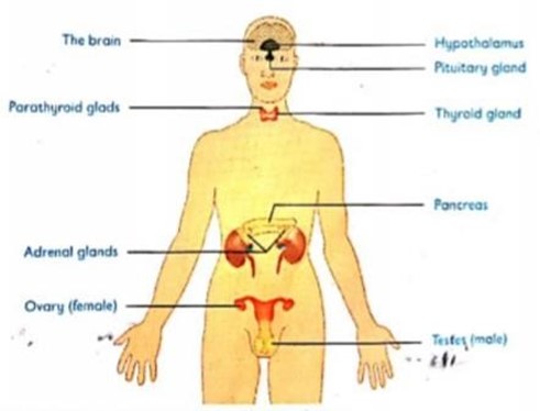
The following are glands/parts of the endocrine system:
i. Pituitary gland
ii. Thyroid gland
iii. Adrenal gland
iv. Parathyroid gland
v. Pancrease
vi. Gonads (Testes and Ovaries)
DIAGRAM OF HUMAN ENDOCRINE SYSTEM
Endocrine system works together with nervous system to help the body to function properly.
SIMILARITIES BETWEEN THE ENDOCRINE SYSTEM AND THE NERVOUS
SYSTEM
i. They both require a stimulus to trigger a certain response.
ii. They both provide a means of communication within the body of an organism.
iii. They both require a transporting medium.
iv. They both bring about responses that enable an organism to survive in an environment.
v. They both work to regulate the activities of cells, tissues, organs and organ systems in relation to internal and external changes.
THE DIFFERENCE BETWEEN NERVOUS SYSTEM AND HORMONAL SYSTEM
| NERVOUS SYSTEM | HORMONAL SYSTEM |
| (i) Electrical impulses are transported by blood. | Hormones are transported by blood. |
| (ii) Response is fast | Response is slow. |
| (iii)Effects are short-lived | Effects are long-lasting. |
| (iv) Usually specific | May affect more than one target organ |
| (v) Electrical signals are sent through nervous impulses | Chemical substances (hormones) evoke a response. |
| (vi) May be voluntary or involuntary | Always involuntary. |
POSITION OF ENDOCRINE GLAND IN HUMAN BODY
1. PITUITARY GLAND
It is located at the base of the fore brain and connected to the hypothalamus by nerve centre.
It has been called the master gland
ROLES OF PITUITARY GLAND
i. Regulates the activities of the other endocrine glands.
ii. It controls the functioning of the body directly by producing its own growth hormone.
iii. It controls the production of Thyroxin hormone in the thyroid gland.
QUESTION: Why the pituitary gland is also known as a master gland?
ANSWER: Because it regulates the activities of the other endocrine glands.
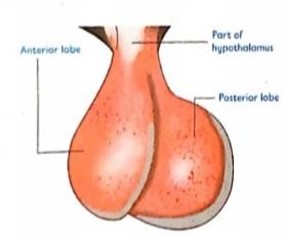
The pituitary gland consists of two different lobes namely:-
i. The anterior lobe
ii. The posterior lobe
Consider the diagram below showing the anterior and posterior lobes
THE ANTERIOR LOBE
This lobe secretes stimulating hormones collectively known as trophic hormones
Anterior lobe also secretes growth hormones and prolactin
TROPHIC HORMONES
Are hormones which activate other endocrine glands to secrete hormones.
Examples of trophic hormones
i. Adrenocorticotrophic hormones (ACTH) – causes the adrenal cortex to secrete cortical hormones
ii. Thyroid stimulating hormones (THS) – causes the thyroid gland to secrete thyroxine hormone.
iii. Gonadotropin hormones e.g. luteinizing hormones (LH) and follicle stimulating hormones (FSH)
Follicle stimulating hormone stimulates the growth of the Graafian follicle in females and causes sperm production in males.
Luteinizing hormone brings about ovulation in female
GROWTH HORMONES
Growth hormones stimulate the body growth.
Effects of growth hormones
i. Over secretion of growth hormones cause gigantism
ii. Under secretion of growth hormones cause dwarfism
PROLACTIN HORMONE
This hormone causes mammary glands to secrete milk in lactating mammals.
THE POSTERIOR LOBE
This lobe secretes antidiuretic hormone and oxytocin
Antidiuretic hormone (ADH) or vasopressin – causes reabsorption of water in the kidneys.
Oxytocin
hormone– brings about contraction of the uterus at birth
Effects of antidiuretic hormone and oxytocin
i. Under secretion of antidiuretic hormone cause Diabetes insipidus.
ii. Hypo secretion of oxytocin cause delayed birth
ii. Over secretion of oxytocin cause premature birth
THYROID GLAND
It is located on the surface of the trachea in the neck
It produces thyroxine hormone
Thryroxine hormone: is an iodine containing hormones. Its function
It rises the metabolic rate by stimulating metabolism of carbohydrates, proteins and lipids
Effects of thyroxine hormone
(i) Under secretion of thyroxine hormone during infancy cause cretinism
(ii) Under-secretion of thyroxine hormone in adult causes myxoedema.
(iii) Deficiency of iodine in the diet cause a disease called Goitre
(iv) Over secretion of thyroxine hormone causes exopthalmic goitre.
CRETINISM
Is a condition characterized by slow physical growth and mental retardation.
MYXOEDEMA
Is a condition characterized by slow physical activity which results in weight gain.
These individuals have a low metabolic rate which is expressed by reduced heartbeat and breathing rate and low body temperature.
GOITRE
Is a disease which is characterized by enlargement of the thyroid gland.
Goitre is an indication of myxoedema in adults.
EXOPTHALMIC GOITRE
The condition is characterized by increase in metabolic rate, causes underweight, restlessness and mental instability.
Also the person becomes thin, excitable and sweats a lot, the eye protrude and the thyroid gland swell.
PARATHYROID GLAND
It is found within the thyroid gland.
It produces parathormone
Parathormone is produced in response to a lack of calcium in the blood resulting increased absorption.
Its function
It controls concentration of calcium and phosphate ions in the blood.
ADRENAL GLANDS
There are two adrenal glands each located above the kidneys.
The adrenal glands consist of an outer layer, the adrenal cortex and an inner layer, the adrenal medulla.
THE ADRENAL CORTEX
Adrenal cortex produces two classes of hormones, namely:
i. Glucocorticoids e.g. cortisol
ii. Mineralocorticoids eg Aldosterone
Cortisol stimulates glucose formation from non-carbohydrates sources e.g. Proteins.
Aldosterone stimulates the reabsorption of sodium ions in the kidney.
Effect of Failure of the adrenal cortex to function properly
Leads to Addison’s disease which is characterized by decrease in blood glucose, loss of sodium chloride and a lot of water in urine.
THE ADRENAL MEDULLA
Adrenal medulla secretes the adrenaline hormone
ADRENALINE HORMONE
Is the hormone for fight or flight
It is produced when an animal is faced with an emergency situation, during anxiety and excitement.
Its function
(i) It prepares the body for a fight or flight action in an emergency
(ii) It prepare the body for emergency by rising blood pressure, increasing heart beat and breathing rates, increasing blood sugar levels and increasing supply of blood to the muscles.
PANCREASE (Islets of Langerhans)
Is a compound gland in that it has both exocrine and endocrine portions.
The exocrine portion produces digestive enzymes.
The endocrine portion produces two hormones namely:
i. Insulin
ii. Glucagon.
Function of insulin
Promotes the conversion of glucose to glycogen by the liver.
Effects of insulin
i. Under secretion of insulin causes Diabetes Mellitus
ii. Over secretion of insulin causes hypoglycaemia
Function of glucagon
Promotes the conversion of glycogen to glucose in the liver cell.
Effects of glucagon
Under secretion of glucagon causes hypoglycaemia
Over secretion of glucagon causes hyperglycaemia
TESTES
They produce male sex hormone collectively known as androgen.
Example of androgen is testosterone hormone
Function of testosterone
i. Regulates the growth, maturation and maintenance of the male reproductive organs.
ii. It is responsible for sperm production
iii. It is responsible for the development of male secondary sexual characteristics. E.g. Beards, deep voice, pubic hairs.
OVARY
Is a female organ which is responsible to produce female sex hormones called oestrogens and progesterones
Function of oestrogen
i. It promotes development of female secondary characteristics. Eg rounded body, growth of breasts
ii. It controls pregnancy by promoting thickening of the uterus
iii. Promotes growth, maturation and maintenance of the female reproductive tract.
Function of progesterone
i. It controls the menstrual cycle
ii. It supports pregnancy
iii. It encourages the development of the uterus lining after ovulation.
iv. It inhibits ovulation and prevents the uterus from contracting during pregnancy.
v. Relaxing is also produced by ovaries begins as the time of birth approaches. This hormone causes the ligaments between the pelvic bones to loosen providing a more flexible passage for the baby during birth
COORDINATION IN PLANTS
Plants perceive and respond to a variety of stimuli that are important to their survival.
Most plant responses are very slow and unnoticeable and normally growth movement
Types of plant responses
i. Tropic response
ii. Tactic response
I. TROPIC RESPONSE
Is the growth movement that is caused by a wide range of stimuli.
Plant grows either towards or away from the stimulus
— If the movement is towards the stimulus, it is called positive tropism.
— If the movement is away from the stimulus, it is called negative tropism.
Tropic movements are mediated through plant hormones
II. PLANT HORMONES
Plant responses are controlled by the following hormones
i. Auxins
ii. Gibberellins
iii. Cytokinins
iv. Ethylene
v. Abscisic acid
AUXINS
Are a group of plant hormones that influence growth
The main auxin made in plants is the indole acetic acid (IAA)
Auxins are produced in buds, root and shoot tips, young leaves, seeds, embryos and developing fruits.
These hormones play an important role in plant tropism.
ROLES OF AUXINS
i. Promote cell elongation.
ii. Promote differentiation of vascular tissues.
iii. Enhance the formation and growth of adventitious roots. This is the reason why stem cuttings grow roots after being put in water containers or soil.
iv. Inhabits development of side branches leading to apical dominance.
v. Promotes formation fruits and elongation of young leaves.
vi. High concentration of auxins stimulates growth of the shoot but inhibits root growth.
NB: Apical dominance is a condition where some plants grow very tall but with few branches.
This can be avoided by cutting the apex of such plants, thus allowing growth of the branches.
EFFECTS OF AUXINS CONCENTRATION ON GROWTH
Experiments have revealed that:
i. High concentration of auxins stimulates growth in shoots
ii. Low concentration of auxins stimulates growth in roots
iii. Amount of auxins which stimulate shoot growth, normally inhibit root growth.
TROPISM
Is the growth movement by plant organs in response to a unilateral stimulus.
Direction of the plant organ movement is related to the direction of the stimulus coming from one direction.
TYPES OF TROPISM
The following are types of tropism
1. Phototropism
2. Hydrotropism
3. Geotropism
4. Thigmotropism
5. Chemotropism
THE IMPORTANCE OF TROPIC AND NASTIC RESPONSES
PHOTOTROPISM
Is the plant growth movement in response to light.
Most plant shoots are said to be positively phototropic because they tend to grow towards light.
Most roots are negatively phototropic because they grow away from light.
Importance of phototropism to plants
i. It places the leaves in a position to maximize light absorption, thereby enhancing photosynthesis.
ii. Etiolation enables seedlings germinating in dark areas to grow fast and reach for light before exhausting their stored food reserves.
EFFECTS OF AUXINS ON PHOTOTROPISM
In an experiment it was revealed that:
1. If a shoot is exposed to light from one direction only, the shoot bends towards the source of light because light detected at the shoot apex causes unequal distribution of auxins. Light causes auxins to migrate to the dark side. Auxins on the dark side become more concentrated than on the side where the light is coming from. The cells on the dark side grow faster and elongate than the ones on the side where the light is coming from. As a result, the shoot bends towards light
NB: Light stimulus is detected at the shoot apex because of the presence of auxins.
2. If a shoot is exposed to the place where there is equal distribution of light, it grows straight upwards because auxins produced at the shoot apex migrate uniformly down the shoot. Hence, promote equal growth rate of ` 8all cells in the zone of elongation, bringing about normal increase in the height of the shoot.
3. If a shoot whose apex has been cut or covered with aluminium foil or opaque cap is exposed to unilateral light, the shoot grows straight upwards because shoot whose have been cut or covered does not respond to light.
4. Plants grow taller and faster in the dark because auxins are not destroyed by light.
5. Plants which have long weak stems, small leaves and lack chlorophyll is said to be etiolated
Etiolation: is a condition resulted from a plant growing in insufficient light, and is yellow, thin and taller than normal.
THE TABLE BELOW DEMONSTRATING EFFECT OF LIGHT ON A PLANT SHOOT
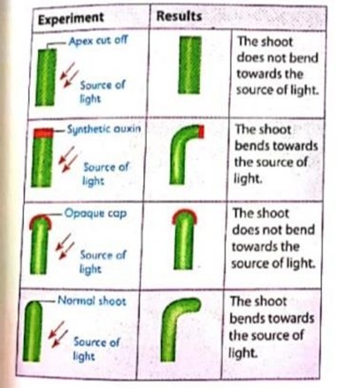
Question: Why plants grow taller and faster in the dark area? Answer: Because auxins are not destroyed by light.
GEOTROPISM
Is the growth movement in response to the force of gravity.
Most roots grow towards the direction of the force of gravity and are said to be positively geotropic.
Most shoots grow away from the force of gravity and are said to be negatively geotropic.
Importance of geotropism to plants
i. It enables plant roots to absorb water and mineral salts from the soil.
ii. It ensures anchorage of the plant in soil to prevent it from falling over or being swept away.
iii. It enables the shoot to grow upwards in order to exposes the plant leaves to sufficient light for photosynthesis.
EFFECTS OF AUXINS ON GEOTROPISM
In an experiment it was revealed that:
1. When a seedling is placed horizontally, auxins accumulate on the lower side due to the force of gravity.
2. The lower side of the root and shoot have more auxins than the upper side,
3. High concentrations of auxins stimulate growth of the shoot but inhibit growth of the root.
4. The lower side of the shoot elongates more than the upper side and the shoot curves upwards.
5. The upper side of the root grows faster than the lower side and the root curve downwards.
Hence, the root is positively geotropic while the shoot is negatively geotropic.
Consider the diagram below showing geotropism response

HYDROTROPISM
Is the growth movement in response to water or moisture.
Roots grow towards moisture and are said to be positively hydrotropic.
When seed planted near a water source such as porous pot or river, the roots of the seedling will always grow towards water.
Auxins at the tip of the root influence growth towards moisture as shown in the figure below:-

IMPORTANCE OF HYDROTROPISM
(i) It enables the plants to absorb dissolved minerals and water.
Water is necessary for various functions such as:
Photosynthesis
Numerous physiological reactions that take place within plant cells.
Turgor pressure, which aids in plant support.
Dissolution of mineral salts.
CHEMOTROPISM
Is the growth movement in response to a unilateral source of chemicals.
Example, during the process of fertilization the pollen tube grows through the style towards the ovule
IMPORTANCE OF CHEMOTROPISM
i. It enables plants to absorb mineral salts from the soil when the roots grow towards beneficial chemicals such as fertilizers.
ii. It facilitates the fertilization process in flowering plants.
THIGMOTROPISM OR HAPTOTROPISM
Is the response of plant organs to the stimulus of touch.
It is mostly exhibited by weak-stemmed plants. Example passion fruits and morning glory
The tendrils of climbing plants bend or twine round a support as a positive response to touch.
The leaves of Mimosa pudica close in response to touch.
Root tips grow away from stones or other obstacles. This is negative haptotropism.
Importance of thigmotropism
1. It helps climbing plants to expose their leaves to sunlight for optimum photosynthesis.
2. Enables plants with weak stems to obtain mechanical support.
3. It enables the insectivorous plants such as the Venus flytrap to trap insects and digest them to obtain nutrients.
EFFECTS OF AUXINS ON THIGMOTROPISM
1. When tendrils or stems of climbing plants come into contact with a suitable hard object, the contact causes them to curve and coil round the object.
2. Contact influences the migration of auxins from the contact surface.
3. The side in contact with object has less auxins so the cells on that side undergo less elongation and therefore less growth.
4. The outer side away from the point of contact has a higher concentration of auxins, promoting faster growth.
5. This causes the shoot to continue coiling round the object.
Consider the diagram below showing a climbing plant coiled around a support

NASTIC MOVEMENT
These are non-directional movement of plant organs in response to diffuse stimuli
Nastic movements are independent of external stimuli
Nastic movements occur as a result of changes in turgor pressure in a certain cells.
Example of nastic movement
i. Folding of leaves in warm weather conditions
ii. Opening and closing of flowers in response to intensity of light. iii. Closing of leaves when touched
TYPES OF NASTIC RESPONSE
i. Nyctinasty or thermometry – is a plant movement in response to temperature changes.
ii. Photonasty – is the plant movement in response to light intensity changes.
iii. Seismonasty – is the plant movement in response to shock or vibrations
iv. Hydronasty – is the plant movement in response to changes in atmospheric humidity.
v. Haptonasty – is the plant movement in response to contact. E.g. Mimosa pudica respond to touch
vi. Chemosnasty – is the plant movement in response to chemicals
TACTIC MOVEMENT
Is the movement of a whole organism in response to an external directional stimulus.
Tactic movements are known as taxis
TYPES OF TACTIC RESPONSES
i. Phototaxis – is the locomotary response to light
ii. Chemotaxis – is the locomotary response to chemicals
iii. Aerotaxis – is the locomotary response to variations in oxygen concentration
iv. Rheotaxis – is the locomotary response to direction of water currents
v. Magnetotaxis – is the locomotary response to magnetic field
vi. Thermotaxis – is the locomotary response to temperature changes
vii. Osmotaxis – is the locomotary response to variations in osmotic pressure
OTHER PLANT HORMONES
GIBBERELLINS
These are mixture of chemical compounds which have an effect on plant growth.
Example of gibberellins
Gibberellic acid
Roles of gibberellins
i. Promotes cell elongation and differentiation
ii. Promotes fruit formation and growth
iii. Breaks seed dormancy
iv. They stimulate rapid growth in dwarf varieties of a certain plants.
CYTOKININS
These are active growth substances which promote growth in plants in the presence of auxins.
They are widely distributed within plants especially in roots.
Roles of cytokinins
i. Promote cell division by inducing growth of roots.
ii. They break seed dormancy
iii. Stimulate opening of stomata
iv. Promote cell enlargement
ETHYLENE
Is a gaseous hormone and the only hormone in gaseous state in plants.
It is produced by fruits when it is about to ripen.
Roles of ethylene
i. Promotes ripening of fruits
ii. Inhibits stem growth
iii. Break bud dormancy
ABSCISIC ACID
Is a hormone which inhibits growth in plants Roles of abscisic acid
i. It promotes falling off of leaves and fruits a process called abscission
ii. Promotes aging
iii. When applied to seeds causes natural dormancy in seeds.
REVISION QUESTIONS
1. An experiment was set up a shown below:
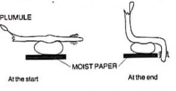
i. Suggest a possible aim of this experiment
a. Plumule
b. Radicle
iii. Account for the response in b (i) and (ii)
2. The diagram below shows a stem of a plant growing round a tree trunk

State the name given to this type of response
3. The diagrams below represents three seedlings grown in a dark chamber with unilateral light source
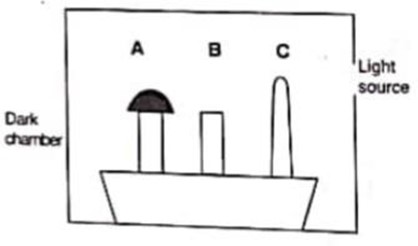
A had the shoot covered with a tin cup while B had the apex cut off and C was left intact. State the results on shoots A, B and C by the end of the experiment and give reasons for your answer
4. Explain the following observations
i. Lighting a shoot from one side makes it bend towards light.
ii. The shoot of a seedling kept in dark placed sideways (horizontal) grows upwards while its roots grow downwards.

